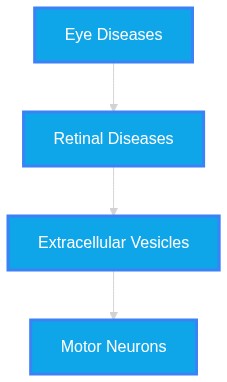
Even though this fact check looked at unique up-to-date abstracts, new evidence may refute this answer in the future. Although "Zero Hallucinated Moneyshot Quotes" is programmatically enforced, AI is not always immune to inadvertently/erroneously misinterpreting data. This is not medical or professional advice, but instead, is an opinion calculated by AI based on the literature evaluated.
"A Top-Down Mechanism for Sporadic ALS Initiated by Ocular Metal Dyshomeostasis and Retrograde Exosomal Transport along the Subcortical Visual Axis"
The provided literature supports a "multi-system" pathology for Amyotrophic Lateral Sclerosis (ALS), which involves both ocular and central nervous system (CNS) components. While there is evidence of visual system involvement—including retinal layer thinning and potential retrograde axonal transport of pathogenic species—a strictly "top-down" ocular-initiated mechanism for sporadic ALS remains a theoretical framework rather than a confirmed biological fact. Evidence confirms that the retina is affected in ALS and could serve as a biomarker, but the precise causality originating specifically from ocular metal dyshomeostasis to systemic motor neuron degeneration is currently underspecified by the literature.
ALS is increasingly redefined as a systemic multi-organ pathology involving both motor and extra-motor neurodegeneration. Ocular and retinal manifestations, specifically retinal ganglion cell layer thinning and microvascular alterations, have been identified as measurable phenotypes. The hypothesis of ocular-initiated disease relies on the known, albeit complex, role of metal ion dyshomeostasis and the retrograde transmission of pathological cargo (e.g., misfolded proteins, aberrant RNA) via extracellular vesicles (EVs). This synthesis evaluates the mechanistic potential for a subcortical visual axis as a primary site of initial pathology.
The traditional view of ALS as a motor-system-restricted disease is challenged by evidence demonstrating widespread ocular and neurovascular dysfunction. Ocular manifestations, including thinning of the retinal nerve fiber layer (RNFL) and ganglion cell layer (GCL), are significantly correlated with functional ALS outcomes. The pathophysiology involves a complex interplay between metal dyshomeostasis—specifically copper and iron—and the propagation of misfolded proteins like TDP-43 or SOD1 through intercellular communication channels. Extracellular vesicles (EVs) have emerged as primary vectors for this propagation. The literature suggests that these vesicles can traverse the blood-brain barrier (BBB) and potentially mediate crosstalk between the retina and the CNS. However, while metal ions like copper and iron are clearly involved in protein misfolding, the evidence to confirm that ocular dyshomeostasis
initiates the systemic disease is currently circumstantial, relying on studies of shared signaling pathways rather than longitudinal proof of origin.
* RNFL thinning is a reproducible biomarker for ALS, suggesting the retina may be an "accessible window" to systemic neurodegeneration.
* Retinal thinning in ALS occurs even in the absence of significant motor symptoms, potentially pre-dating symptomatic onset.
* Metal dyshomeostasis is a central, multifaceted driver in ALS, where free copper and iron imbalances initiate protein aggregation (e.g., SOD1).
* EVs released from muscle, glial, and neuronal cells act as "messengers" that spread pathogenic proteins and miRNAs, potentially reinforcing a feedback loop of systemic dysfunction.
* The retina and visual pathways exhibit structural damage such as demyelination and thinning of RGCs, which shares neuroimmune features with cerebral pathology.
* Copper and iron levels in the spinal cord are disrupted in sporadic ALS, parallel to findings in familial cases, suggesting a shared metabolic end-point.
* The use of OCT and OCT-A allows for the non-invasive mapping of vascular and structural neurodegeneration that may eventually function as a clinical diagnostic tool.
1. PubMed ID:
42526365- Application: The text discusses the diagnostic potential of OCT in ALS. "This meta-analysis demonstrates significant bilateral RNFL thinning in ALS, with relative preservation of the Inner Nuclear Layer and Ganglion Cell Layer - Inner Plexiform Layer, supporting retinal neurodegeneration as a feature of this multisystem disorder."
2. PubMed ID:
42512084- Application: The text confirms retinal microvascular alterations. "ALS is associated with structural and microvascular retinal alterations detectable by OCT and OCT-A. These findings support the presence of systemic neurovascular dysfunction and highlight retinal imaging as a promising, non-invasive approach for investigating disease mechanisms and developing potential biomarkers."
3. PubMed ID:
40625857- Application: The text describes inner retinal pathology progression in mouse models. "Our findings demonstrate distinct inner retinal nerve fiber layer pathology, detected using cSLO coupled with OCT, which worsens over time. These findings support the potential of retinal imaging as a translationally relevant, non-invasive biomarker for ALS diagnosis or disease monitoring in humans."
4. PubMed ID:
40306255- Application: The text notes specific layer changes. "The examination of retinal layers in ALS patients revealed a significant change in the inner nuclear layer (INL), with a pattern of initial thickening followed by thinning, which correlated with disease stages, most notably in the inner nasal quadrant."
5. PubMed ID:
40698100- Application: The text highlights GCL thickness correlations. "the significant positive correlation observed between GCIPL and ALS functional outcome and between RNFL and GCIPL measurements highlighted the fact that though the axonal degeneration in retinal neurons might not be translating to the same extent in ganglion cells in ALS, the subtle thinning of GCIPL correlated strongly with functional disability in patients with ALS"
6. PubMed ID:
40560963- Application: The text links retinal layer thickness to ALS risk. "a standard deviation (SD) decrease of 15.19 µm in photoreceptor layer (PRL) thickness was associated with a 19% (95% confidence interval [7, 29]; p = 0.002) increased risk of ALS, while a SD increase of 26.11 µm in retinal pigment epithelium (RPE) thickness corresponded to a 20% (95% CI [7, 34]; p = 0.002) higher risk."
7. PubMed ID:
40350723- Application: The text discusses non-typical oculomotor manifestations. "Oculomotor disorders are not typical manifestations of amyotrophic lateral sclerosis (ALS). Occasionally, this disease is associated with vertical gaze paresis, presenting a distinct type as «ALS+progressive supranuclear palsy»."
8. PubMed ID:
33071739- Application: The text describes systemic ALS involvement. "This pathology affects the motor neurons, the spinal cord, the cerebellum, and the brain, but recently, it has been shown that it also affects the visual system. This impact occurs not only at the level of the oculomotor system but also at the retinal level, which is why the retina is being proposed as a possible biomarker of this pathology."
9. PubMed ID:
40340620- Application: The text discusses ET utility. "By synthesizing findings on the connection between oculomotor dysfunction and cognitive decline, this review underscores the potential of ET as a noninvasive tool for assessing ALS progression."
10. PubMed ID:
38467696- Application: The text discusses spinal cord copper levels in sporadic ALS. "We found that when compared to control cases the natural distribution of spinal cord copper was disrupted in sporadic ALS. A standout feature was decreased copper levels in the ventral grey matter, the primary anatomical site of neuronal loss in ALS."
11. PubMed ID:
39050823- Application: The text discusses copper homeostasis. "The pathophysiology of ALS involves many signs of a disruption in copper homeostasis, with both excess free levels and functional deficiency likely occurring simultaneously."
12. PubMed ID:
33206086- Application: The text notes shared pathway toxicity. "Interestingly, we verified that Mn-induced neurotoxicity shares pathways associated with the development of Alzheimer's disease, Amyotrophic Lateral Sclerosis, Huntington's disease, and Parkinson's disease."
13. PubMed ID:
40894549- Application: The text discusses lead toxicity in the optic nerve. "Our findings suggested that Pb toxicity may impair survival and maturation process of oligodendrocytes, changes in myelin structures, and potential demyelination of the optic nerve."
14. PubMed ID:
41651252- Application: The text discusses EV cargo. "Overall, our findings show that trimeric SOD1 influences EV cargo and spread in ALS."
15. PubMed ID:
42351263- Application: The text discusses SkM-EVs. "In these contexts, SkM-EVs may contribute to disease progression by delivering pathogenic cargo, including misfolded proteins and aberrant RNAs, to motor neurons."
16. PubMed ID:
39955442- Application: The text discusses SMN delivery via EVs. "In this study, we show that adenovirus-mediated overexpression of SMN protein in HepG2 cells leads to the release of EVs loaded with high levels of SMN protein into conditioned medium."
17. PubMed ID:
42156174- Application: The text discusses COMMD1 and SOD1. "In vivo investigations utilizing male hSOD1G93A transgenic mice demonstrated that COMMD1 deficiency markedly ameliorated the deterioration of motor function and prolonged survival duration."
18. PubMed ID:
42097486- Application: The text discusses neuronal death and copper redistribution. "This indicates that following neuronal death, Cu might be expelled from brain proteins and subsequent to redistribution between brain, CSF and blood, it will result a brain Cu deficiency and a decrease in Cu brain protection against free radicals."
19. PubMed ID:
41672113- Application: The text links SOD1 to EV shedding. "Deletion of SOD-1, as well as the SOD-1(G85R) amyotrophic lateral sclerosis (ALS) pathogenic variant, increased EV shedding from the cilium distal tip, and this was associated with greater abundance of EV cargo in this ciliary compartment."
20. PubMed ID:
42304926- Application: The text notes the retina as a brain outgrowth. "Both these structures within the central nervous system (CNS) utilize common neuro-inflammatory mechanisms because the retina is an outgrowth of the brain."