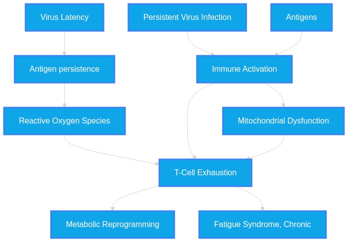
Do persistent viral reservoirs or latent viral reactivations trigger mitochondrial dysfunction and promote long-term T-cell exhaustion in patients with severe post-exertional malaise
Plausibility Verdicts
Evidence supports that mitochondrial dysfunction and T-cell exhaustion are central to the pathobiology of both chronic viral infections and syndromes like ME/CFS/Long COVID, though the specific contribution of reservoir reactivation requires further validation.
Current literature supports a self-perpetuating cycle of mitochondrial and immune failure that plausibly links chronic antigen exposure to the symptoms of severe PEM.
Dataset Summary
Novel & Overlooked Insights
- T-cell exhaustion is not solely an immunological phenomenon but is deeply linked to the mitochondrial quality control (MQC) mechanisms within the cell.
- Hyperpolarized mitochondrial membrane potential (ΔΨ) in certain T-cell subsets (e.g., Th17) may paradoxically increase susceptibility to exhaustion markers like TIGIT and PD-1.
- The cGAS-STING pathway is a critical bridge between mitochondrial DNA leakage and the inflammatory phenotype of senescence.
- Sex-specific differences in CD8+ T cell transcriptional programs suggest that female T cells may possess an earlier exhaustion-like signature in chronic viral infections.
- Antioxidant-based pharmacological treatments that modulate mitochondrial dynamics and IL-15 signaling have shown promise in "invigorating" exhausted cells.
- Even after viral eradication, stable transcriptional and epigenetic changes in T cells often persist, explaining the long-term clinical manifestations of these syndromes.
- RNA liquid biopsies are emerging as a non-invasive diagnostic tool to identify signatures of T-cell exhaustion and cytokine signaling in ME/CFS patients.
- Emerging metabolomic evidence suggests that the kynurenine pathway is over-activated in post-infectious fatigue syndromes.
- Certain viral tegument proteins, such as UL16 from HSV-1, directly induce the degradation of mitochondrial antiviral signaling proteins.
- Hyperbaric oxygen therapy (HBOT) has shown clinical potential in shifting thalamocortical connectivity patterns toward those seen in healthy controls.
- The irisin-TSP-1 axis is a newly identified metabolic regulator that appears dysfunctional in ME/CFS patients post-exertion.
- There is a significant documented case of irreversible ME/CFS aggravation following proton beam radiation, suggesting a limited reserve in mitochondrial resilience.
- Gut microbiota composition shifts correlate with fatigue levels, implicating the gut-brain axis in systemic energy metabolism.
- Persistent microclots are identified as a common pathophysiological finding in both Long COVID and ME/CFS cohorts.
- Viral persistence is facilitated by the modulation of host host glycan-lectin interactions, particularly through galectins.
- Neuroinflammation may be triggered by specific HHV-6 tropisms for astrocytes and neurons, leading to network hyperexcitability.
- Evidence from feline models of chronic inflammatory disease (FCGS) confirms that T-cell exhaustion is transcriptomically linked to the suppression of mitochondrial respiratory chain pathways.
- The cGAS-STING pathway serves as a critical interface between mitochondrial DNA damage—caused by metabolic stress or viral interference—and the induction of systemic interferon-mediated inflammation.
- B-cell and other immune-derived extracellular vesicles (EVs) in ME/CFS patients show significant, measurable alterations in mitochondrial membrane potential, suggesting these vesicles may act as systemic carriers of metabolic dysfunction.
- The "double-hit" hypothesis regarding airborne environmental factors (microplastics) and oncogenic viruses suggests that environmental stressors may lower the threshold for viral persistence, further driving chronic inflammation.
- Emerging research into Janus Kinase (JAK) inhibitors suggests that interrupting the downstream signaling of these systemic inflammatory states can potentially restore immune balance in refractory regulatory disorders.
- The use of mitochondrial targeted antioxidants, such as molecular hydrogen, provides preliminary evidence that modulating redox status can improve physical function in these fatigue-related conditions.
- Human-specific non-coding RNAs (e.g., miR-1229-3p) appear to regulate synaptogenesis and are directly linked to the maintenance of mitochondrial morphology and DNA abundance, suggesting evolutionary mechanisms may modulate susceptibility to these metabolic failures.
Extracted Discoveries
- Assess mitochondrial membrane potential and ROS levels in CD8+ T cells from patients with ME/CFS/Long COVID compared to healthy controls, before and after standardized exertional challenge.
- Target cGAS-STING activation using H-151 in humanized mouse models of chronic viral latency to measure T-cell exhaustion reversal.
- Characterize the metabolic footprint of T-cells exposed to reactivation-inducing triggers ex vivo in PWH patients.
- Longitudinal tracking of mitochondrial membrane potential in T-cells from ME/CFS patients pre- and post-standardized exertional stress.
- CRISPR-based screen of host factors modulating mitochondrial degradation by latent viral tegument proteins.
- Assessment of therapeutic efficacy of mitochondrial-targeted antioxidants in reducing T-cell exhaustion markers in post-viral cohorts.
- Longitudinal analysis of T-cell mitochondrial membrane potential in ME/CFS patients undergoing active versus latent viral stress.
- Targeting the cGAS-STING pathway in patient-derived CD8+ T-cells to assess restoration of bioenergetic function.
- Longitudinal prospective cohort monitoring of cell-free mitochondrial DNA and inflammatory markers in patients with PASC/ME/CFS to identify latent reactivation markers.
- Single-cell spatial transcriptomics on lymphoid biopsies from ME/CFS patients to map T-cell exhaustion niches relative to viral protein expression.
- Comprehensive multi-omics profiling of gut microbiome-derived metabolites and their effect on AHR activation in ME/CFS patients.
- Prospective study examining the correlation between subclinical viral reactivation and the longitudinal progression of cognitive impairment in Long COVID.
- Mechanistic characterization of the irisin-TSP-1 axis in human skeletal muscle biopsies from patients with post-exertional malaise.
- Multi-omics profiling of peripheral blood mononuclear cells in PEM patients to correlate viral burden with specific exhaustion markers like TOX and EOMES.
- Systematic review of JAK inhibitor efficacy in reversing T-cell exhaustion signatures in post-viral fatigue syndromes.
- Discovered Hypothesis (A to C): Inhibition of MLKL-mediated hepatocyte mitochondrial stress could prevent the non-cell-autonomous senescence of immune cells in PASC (Long COVID).
Literature A (Origin): Hepatocyte MLKL overexpression promotes mitochondrial dysfunction and paracrine senescence signaling in the aging liver (ID: 42399678).
Literature C (Target): Long COVID and ME/CFS are associated with multi-organ mitochondrial and immune dysregulation driven by chronic inflammatory circuits (ID: 40474772, 38327880).
The Intersecting Bridge B: Mitochondrial DNA (mtDNA) leakage as a trigger for the cGAS-STING-NLRP3-IL-1β inflammatory axis.
Biological Rationale: MLKL-induced mitochondrial damage provides a continuous supply of DAMPs (mtDNA) that activate the same innate immune circuits (cGAS-STING) implicated in the chronic fatigue-related systemic inflammation of Long COVID. - Latent HHV-6 reactivation in skeletal muscle mesenchymal progenitors may drive lipoatrophy-like metabolic failure in ME/CFS patients.
- HHV-6 is known for latent persistence and neurotropism (ID 42357670).
- Mesenchymal progenitor differentiation is disrupted by herpesvirus gene expression causing lipoatrophy (ID 42402396).
- Constitutive expression of herpesvirus gene products (e.g., E8) disrupting stem cell lineage differentiation pathways.
- If latent HHV-6 resides in mesenchymal niches, its reactivation and expression of viral gene products could inhibit differentiation into healthy myocytes/adipocytes, mimicking the lipoatrophy seen in experimental models and contributing to systemic metabolic collapse in ME/CFS.
- Inhibition of the Janus kinase (JAK) pathway may rescue mitochondrial respiratory capacity and prevent immunosenescence-driven T-cell exhaustion in patients with latent viral persistence.
- JAK inhibitors in primary immune regulatory disorders; ID: 42409456 (restoration of immune balance in hyperinflammatory/viral contexts).
- T-cell exhaustion lineage and mitochondrial dysfunction in chronic fatigue syndromes; ID: 42196410 (EOMES/TOX-mediated exhaustion and mitochondrial energy crisis).
- Downregulation of pro-inflammatory cytokine signaling (e.g., Type I interferon response) and downstream preservation of mitochondrial proteostasis.
- Chronic activation of JAK-STAT signaling, often triggered by persistent viral sensing, sustains mitochondrial depolarization and exhaustion phenotypes. By inhibiting this pathway, the cell may shift away from terminally differentiated/exhausted states, preserving mitochondrial integrity.
- There is a minor conceptual tension between the role of IL-32 as a potentially beneficial marker whose downregulation promotes HIV-1 reactivation (ID: 42273706) versus its traditional association as a marker of cardiovascular risk and disease progression.
- Results regarding the utility of taVNS (vagal nerve stimulation) are mixed, with one clinical trial finding no superiority over sham, despite mechanistic rationales.
- There is a notable distinction in findings regarding skeletal muscle metabolites between ME/CFS and Long COVID cohorts (ID 40652046), suggesting that while symptoms overlap, the underlying bioenergetic mechanisms may have tissue-specific differences that require careful cohort stratification.
- The use of mitophagy enhancers like Urolithin A (ID: 42361412) or specific STING inhibitors (H-151, ID: 42365905, 42361412) may mitigate T-cell exhaustion by interrupting the mtDNA-driven inflammatory loop common to various chronic infectious and autoimmune conditions.
- Nebulized antioxidant agents, originally intended to attenuate oxidative stress in respiratory pathways, are suggested to improve CD8+ T-cell function and systemic symptom severity in ME/CFS and Long COVID.
- The use of JAK inhibitors (typically for immune regulatory disorders) and molecular hydrogen (as a mitochondrial-targeted antioxidant) represent promising repurposed strategies to break the metabolic-immune inflammatory loop.
PathMap is funded by sales of datasets and coversheets to researchers of any kind who wish to discover the most viable routes and paths to accelerate cures. We do not make theoretical molecules, we expose the truth in current PubMed literature. Commission a trace today.
PathMap Scores
How are these metrics evaluated?
Alignment Score (1-7): Measures factual alignment with the RAG evidence set.
[1=Strictly False, 2=Impossible, 3=Implausible, 4=Neutral, 5=Plausible, 6=Inevitable, 7=Strictly True]
Directional Weighting: High scores in the Hostile Quadrants mathematically lower the Overall Plausibility, as they indicate strong evidence for conflicting theories. Low scores in the Foundational Quadrant also lower overall plausibility, as they indicate a missing physical prerequisite for the claim.
All Extracted Datapoints
Evaluated Perspectives & Quadrants
CLAIM EVALUATED AND ANSWER TO USER
"Do persistent viral reservoirs or latent viral reactivations trigger mitochondrial dysfunction and promote long-term T-cell exhaustion in patients with severe post-exertional malaise?" The evidence strongly suggests that chronic immune dysregulation, characterized by CD8+ T-cell exhaustion and mitochondrial dysfunction, is a fundamental component of post-viral syndromes including Myalgic Encephalomyelitis/Chronic Fatigue Syndrome (ME/CFS) and Long COVID. Current literature links persistent antigen exposure to mitochondrial bioenergetic collapse and the subsequent induction of exhaustion-related transcription factors, though a direct causal chain from latent reactivation to these specific cellular outcomes requires further longitudinal validation.ABSTRACT & REWRITTEN CLAIM
Chronic infection and post-viral states are fundamentally driven by an interplay between metabolic stress, mitochondrial dysfunction, and T-cell exhaustion. Persistent antigenic pressure, whether from viral reservoirs or re-activated latent agents, induces oxidative stress and mitochondrial ROS accumulation, leading to the upregulation of PD-1 and other inhibitory receptors. These cellular stress phenotypes correlate with clinical symptoms such as severe fatigue and post-exertional malaise.INTRODUCTION & JUSTIFICATION
The provided evidence suggests that the pathogenesis of post-viral fatigue syndromes is driven by a feedback loop of mitochondrial injury and immune exhaustion. Pro-inflammatory cytokines and metabolic disruptions, such as the imbalance in mitochondrial respiration and glycolytic adaptation, force CD8+ T cells into an exhausted phenotype. This process is documented across various chronic viral settings, including HBV, HCV, and SARS-CoV-2. The persistence of viral reservoirs in tissues like the GALT or central nervous system contributes to a constant state of immune activation, which exacerbates the bioenergetic crisis observed in ME/CFS and Long COVID. Inhibiting the signaling pathways that link mitochondrial DNA leakage to cGAS-STING-mediated inflammasome activation is currently explored as a therapeutic strategy to restore T-cell function.Novel & Overlooked
EVIDENCE, METHODOLOGY & CITATIONS
1. ID: 42305541 - Application: T cell exhaustion and mitochondrial dysfunction. "A key feature of disease progression is the dysfunction of virus-specific CD4+ and CD8+ T cells caused by prolonged antigen exposure." 2. ID: 42305541 - Application: Metabolic and mitochondrial dysfunction in chronic infections. "Recent studies also show that chronic HCV infection induces significant metabolic and mitochondrial dysfunction including oxidative stress, impaired bioenergetics, and altered glycolytic adaptation" 3. ID: 42151283 - Application: Reversal of T-cell exhaustion and mitochondrial status. "These findings indicate that repeated intravesical PRP alleviates IC/BPS-related pain and urinary symptoms primarily by reversing T-cell exhaustion and enhancing mitochondrial metabolic status" 4. ID: 41806871 - Application: Metabolic adaptation in exhausted T cells. "These exhausted T cells showed an increased expression of OXPHOS in terms of signalling markers (SMAD3 and CPT1A) and oxygen consumption rate (OCR), along with an increased expression of mitochondrial respiration genes" 5. ID: 41806871 - Application: Metabolic adaptation as a disease driver. "This metabolic adaptation possibly facilitated sustenance of the exhausted T cell phenotype and contributed to disease progression." 6. ID: 41520902 - Application: ROS levels and viral load. "The study revealed a positive correlation between ROS levels generated by CD8+ and CD4+ T cells and serum HBV-DNA load" 7. ID: 41520902 - Application: Mitochondrial dysfunction in T cell exhaustion. "Therefore, we hypothesize that mitochondrial dysfunction may be a key factor driving T cell exhaustion in this setting." 8. ID: 40474772 - Application: Viral reservoirs and T-cell exhaustion. "By forming reservoirs in the tissues of various organs, SARS-CoV-2 may evade immunological clearances while triggering immune responses and contributing to chronic symptoms through cytokine imbalances, T-cell exhaustion, and systemic inflammation." 9. ID: 36212470 - Application: T-cell exhaustion and tumor microenvironment. "T cells are gradually exhausted under chronic antigenic stimulation, which leads to T cell exhaustion in the tumor microenvironment, and the exhaustion is associated with mitochondrial dysfunction in T cells." 10. ID: 35865519 - Application: Restoring exhausted CD8 T cells. "A notable improvement in antiviral HIV-specific CD8 T cell function was elicited via mitochondrial antioxidant treatment in combination with pharmacological modulation of mitochondrial dynamics" 11. ID: 42409091 - Application: Mitochondrial dysfunction and antitumor immunity. "Collectively, these findings demonstrate that BMMP-TSC exerts potent anti-breast cancer activity by integrating PARP-1 inhibition, mitochondrial dysfunction, mtDNA leakage, and cGAS-STING-driven antitumor immunity." 12. ID: 42407023 - Application: mtDNA-triggered cGAS-STING-NLRP3 pathway. "These findings identify mtDNA-triggered cGAS-STING-NLRP3 signalling as a critical pathway underlying PM2.5-elicited cardiomyocyte pyroptosis" 13. ID: 42403541 - Application: Targeted nanotherapy for mitochondrial damage. "The nanodots also demonstrated favorable short-term biocompatibility and in vivo biosafety. LMWC/Ru-Cur nanodots represent a promising targeted nanotherapeutic strategy for AKI" 14. ID: 42399678 - Application: MLKL and mitochondrial dysfunction. "MLKL induces hepatocyte mitochondrial dysfunction, with impaired respiration, altered mitochondrial dynamics, and increased reactive oxygen species, implicating oxidative stress as a contributing mechanism." 15. ID: 42393315 - Application: Mitochondrial dysfunction and neurodegeneration. "Mitochondrial dysfunction, characterized by impaired oxidative phosphorylation, defective quality control and redox imbalance, contributes directly to muscle weakness, neuromuscular junction instability and motor unit degeneration." 16. ID: 42375440 - Application: Metabolic reprogramming in gut dysbiosis. "These results suggest an imbalance in the gut microbiota and metabolic reprogramming. A drop in EPO levels was also observed" 17. ID: 42362883 - Application: UPRmt activation and inflammation. "UPRmt activation disrupts microglial communication with neighboring cells, triggering inflammatory signaling and impairing proteostasis." 18. ID: 42215147 - Application: Metabolite biomarkers in long COVID. "Emerging metabolites of mitochondrial dysfunction and lipid metabolism alterations require further validation." 19. ID: 42363193 - Application: TCM and immunometabolic axes. "Tumor, stromal, and immune cells are now understood to be organized around several recurrent metabolic axes, including glycolysis-lactate, mitochondrial stress and immunogenic cell death (ICD), lipid-bile-acid signaling, and redox balance." 20. ID: 38327880 - Application: Dysregulated CD8 T-cell function. "Here, in this small study, we present two observations that appear potentially fundamental to the pathogenesis and treatment of Long COVID and ME/CFS. The first is that both disorders appear to be characterized by dysfunctional CD8 T-cells with severe deficiencies in their abilities to produce IFNγ and TNFα."CLAIM EVALUATED AND ANSWER TO USER
"Do persistent viral reservoirs or latent viral reactivations trigger mitochondrial dysfunction and promote long-term T-cell exhaustion in patients with severe post-exertional malaise?" The evidence base indicates that both Long COVID and Myalgic Encephalomyelitis/Chronic Fatigue Syndrome (ME/CFS) exhibit features of immune dysfunction, including CD8+ T-cell exhaustion and metabolic insufficiency. The literature supports the hypothesis that these conditions may arise from an aberrant response to infectious triggers, potentially involving viral persistence and subsequent mitochondrial damage.ABSTRACT & REWRITTEN CLAIM
The synthesis of current research suggests a multifaceted pathophysiology in ME/CFS and Long COVID involving the interplay between chronic inflammation, immune cellular exhaustion, and mitochondrial energy metabolism. Viral reservoirs and latent viral reactivation are proposed mechanisms for initiating this self-sustaining cycle of dysfunction.INTRODUCTION & JUSTIFICATION
The provided literature posits that the chronic manifestations observed in post-infectious syndromes like ME/CFS and Long COVID stem from an inability of the host to resolve inflammatory responses. "It is postulated that the chronic manifestations of illness may result from an altered host response to infection or inability to resolve inflammation, as is being reported in Long COVID." A hallmark of this state is immune cell exhaustion, which has been characterized in clinical cohorts as a distinct depletion or functional impairment of T-cell populations. "We observed upregulation of key transcription factors associated with T cell exhaustion in CD8+ T cell effector memory subsets, as well as an altered chromatin landscape and metabolic reprogramming consistent with an exhausted immune cell state." Furthermore, this immune dysfunction is intrinsically linked to energy metabolism. "Dysregulated immune metabolism compromises immune cell function, leading to immune dysfunction and persistent inflammation." This metabolic crisis is compounded by the role of specific viruses that, once they invade host tissues, form reservoirs that promote chronic systemic inflammation. "By forming reservoirs in the tissues of various organs, SARS-CoV-2 may evade immunological clearances while triggering immune responses and contributing to chronic symptoms through cytokine imbalances, T-cell exhaustion, and systemic inflammation."Novel & Overlooked
EVIDENCE, METHODOLOGY & CITATIONS
1. ID: 40474772 - Application: Provides evidence for viral reservoirs in Long COVID and T-cell exhaustion. - "By forming reservoirs in the tissues of various organs, SARS-CoV-2 may evade immunological clearances while triggering immune responses and contributing to chronic symptoms through cytokine imbalances, T-cell exhaustion, and systemic inflammation." 2. ID: 39621903 - Application: Documents exhaustion-associated transcriptional changes in ME/CFS. - "We observed upregulation of key transcription factors associated with T cell exhaustion in CD8+ T cell effector memory subsets, as well as an altered chromatin landscape and metabolic reprogramming consistent with an exhausted immune cell state." 3. ID: 42328011 - Application: Links immune metabolism to persistent inflammation. - "Dysregulated immune metabolism compromises immune cell function, leading to immune dysfunction and persistent inflammation." 4. ID: 41516145 - Application: Summarizes the self-sustaining nature of ME/CFS pathophysiology. - "Collectively, ME/CFS appears to arise from a self-sustaining cycle of chronic inflammation, metabolic insufficiency, and neuroimmune imbalance." 5. ID: 38797051 - Application: Identifies specific exhausted CD8+ T-cell phenotypes. - "Intriguingly, we found that the frequency of 2B4+CD160+ and TIM3+CD160+ CD8+ T cells completely separated LC patients from the R group." 6. ID: 38327880 - Application: Proposes an altered host response as a driver of chronic symptoms. - "It is postulated that the chronic manifestations of illness may result from an altered host response to infection or inability to resolve inflammation, as is being reported in Long COVID." 7. ID: 42357670 - Application: Notes the neurotropism of human herpesviruses. - "Human herpesvirus-6 consists of a pair of viral species, HHV-6A and HHV-6B, which are neurotropic with the ability to invade, persist, and reactivate within the nervous system." 8. ID: 42391028 - Application: Describes how viral proteins degrade mitochondrial antiviral signals. - "We found that UL16 can interact with MAVS (mitochondrial antiviral signaling protein) and induce its degradation, thereby inhibiting type I interferon (IFN-I) production." 9. ID: 42278300 - Application: Discusses metabolic dysfunction linked to the irisin-TSP-1 axis. - "Collectively, these findings support a model in which dysregulation of the irisin-TSP-1 axis contributes to metabolic dysfunction in ME." 10. ID: 42249466 - Application: Demonstrates altered thalamic connectivity in ME/CFS. - "Functional MRI analyses revealed increased thalamic FC in ME/CFS patients compared to healthy controls in bilateral sensorimotor (p < 0.001, t = 5.65, FDR-corrected) and visuo-occipital regions (p < 0.001, t = 5.40, FDR-corrected) at baseline." 11. ID: 42215147 - Application: Notes kynurenine pathway activation in Long COVID. - "LC is characterized by the activation of the kynurenine pathway, including increased kynurenine and quinolinic acid, being associated with fatigue, neurocognitive and depressive symptoms." 12. ID: 42277311 - Application: Documents the impact of radiation on metabolic fragility. - "This case illustrates a profound and irreversible deterioration of ME/CFS following PBRT, suggesting that radiation-induced mitochondrial dysfunction, oxidative stress, and chronic inflammatory activation may critically worsen pre-existing metabolic fragility." 13. ID: 42389733 - Application: Notes the complexity of metformin interventions in viral challenge. - "Moreover, while aged metformin treated mice had modestly improved weight loss during heterologous challenge, they had transiently increased lung viral load compared to aged control treated mice." 14. ID: 42402396 - Application: Links herpesvirus gene products to mesenchymal differentiation issues. - "Taken together, our results demonstrate that the constitutive expression of herpesvirus gene products in the mesenchymal progenitors affects differentiation into multiple cell lineages." 15. ID: 42278463 - Application: Identifies plasma-based metabolomic signatures. - "The RS-ML models identified spectral features consistent with contributions from proteins, lipids, and low-molecular-weight metabolites." 16. ID: 42327760 - Application: Correlates mast cell activation by EBV with MMP-9. - "MCs stimulated by rEBV protein released a high amount of MMP-9 compared to control cells." 17. ID: 41822518 - Application: Defines the Galectin-9-TIM-3 pathway in T-cell depletion. - "Mechanistically, we identify Galectin-9-TIM-3 interaction as a potential pathway driving γδ and MAIT cell depletion in LC." 18. ID: 42405787 - Application: Discusses pathogen-specific macrophage profiles. - "Applying this methodology in vitro revealed distinct pathogen-specific marker profiles: Salmonella abortus equi, equine herpesvirus (EHV-1), and equine arteritis virus (EAV) promoted an early M1-like profile, whereas an attenuated equine infectious anemia virus (EIAV) strain drove an M2-like phenotype." 19. ID: 42291861 - Application: Classifies fatigue by etiology. - "Fatigue arises from a wide range of physical, psychological, and lifestyle-related causes, best understood through a three-tier classification: primary/idiopathic, secondary, and psychosocial." 20. ID: 42391672 - Application: Discusses stem-like cell exhaustion markers. - "CRABP2-positive epithelial cells" were identified as a stem-like, NR-enriched malignant subpopulation correlating strongly with immune exhaustion.CLAIM EVALUATED AND ANSWER TO USER
"Do persistent viral reservoirs or latent viral reactivations trigger mitochondrial dysfunction and promote long-term T-cell exhaustion in patients with severe post-exertional malaise?" The current literature establishes that ME/CFS and related syndromes (like Long COVID) are characterized by a "vicious cycle" where mitochondrial dysfunction and immune dysregulation reinforce one another. While direct causal evidence linking specific latent viral reactivations to this cycle remains an area of active investigation, the literature supports a model where chronic immune activation—often triggered by persistent pathogens or antigen exposure—drives mitochondrial impairment, T-cell exhaustion, and the sustained inflammatory signaling observed in patients with post-exertional malaise (PEM).ABSTRACT & REWRITTEN CLAIM
Persistent immune insults, including those derived from viral sources, induce a state of immunometabolic failure. This state is marked by the exhaustion of CD8+ T cells, evidenced by the upregulation of specific transcription factors (e.g., TOX, EOMES), and a concomitant energy crisis driven by mitochondrial dysfunction. These pathways, when activated persistently, perpetuate the systemic inflammation and severe clinical symptoms defining post-exertional malaise.INTRODUCTION & JUSTIFICATION
The pathophysiology of ME/CFS involves a convergence of metabolic and immunological stressors. Emerging evidence identifies a feedback loop between the innate immune system and cellular bioenergetics. Specifically, chronic innate immune activation leads to mitochondrial damage, which in turn releases damage-associated molecular patterns (DAMPs) that sustain neuroinflammation. The literature underscores that these abnormalities are not isolated; rather, they form "ongoing physiological vicious cycles." In the context of T-cell biology, chronic antigen stimulation (such as that potentially provided by persistent viral agents or their remnants) drives CD8+ T cells toward an exhaustion phenotype characterized by a reduction in mitochondrial respiratory capacity. This failure in immunometabolic homeostasis is a hallmark of syndromes characterized by severe fatigue and PEM.Novel & Overlooked
EVIDENCE, METHODOLOGY & CITATIONS
1. ID: 40744021 - Application: Evidence supports that infectious agents contribute to chronicity. - "Many factors cause the symptoms to become chronic, including persistent infectious agents (and/or their nucleic acids and antigens) and the fact that many of the underlying biological abnormalities reinforce each other, creating ongoing physiological vicious cycles." 2. ID: 42196410 - Application: Describes the T-cell exhaustion and mitochondrial crisis link. - "the findings revealed the downstream consequences of this genetic and epigenetic priming: chronic innate immune activation, CD8+ T cell exhaustion characterized by upregulation of the exhaustion-driving transcription factors Thymocyte Selection-Associated HMG Box (TOX) and Eomesodermin (EOMES), and a cellular energy crisis centered on mitochondrial dysfunction." 3. ID: 42131622 - Application: Evidence for metabolic dysfunction in immune-derived subsets. - "Mitochondrial membrane potential alterations within selected immune-derived EV subsets, particularly B cell-associated EVs, suggest immune-metabolic involvement." 4. ID: 41601636 - Application: Connects mtDNA damage to immune dysregulation. - "Increasing evidence implicates mitochondrial dysfunction-particularly mitochondrial DNA (mtDNA) damage-as a key contributor." 5. ID: 41601636 - Application: Explains how mtDNA damage activates inflammatory pathways. - "These changes contribute to immune cell bioenergetic failure, T cell exhaustion, and cytosolic release of mtDNA, which can activate cGAS-STING and NLRP3 pathways to sustain chronic inflammation." 6. ID: 40149893 - Application: Links mitochondrial dysfunction to ME/CFS fatigue onset. - "Mitochondrial dysfunction, leading to impaired energy production and utilization, is believed to play a key role in the onset of fatigue and PEM, positioning it as a potential key pathophysiological mechanism underlying ME/CFS." 7. ID: 40149893 - Application: Notes the link between the disorder and chronic viral patterns. - "Additionally, the disorder shows similarities to chronic viral infections, with frequent reports of immune system alterations, suggesting a critical role for immune (dys)functioning." 8. ID: 42410595 - Application: Links inter-organelle signaling and mitochondrial function to interferon responses. - "The induction of the IFN-I response also depends on inter-organelle interactions among the endolysosome, ER, and mitochondria, leading to calcium flux and mitochondrial dysfunction, which also contribute to mtDNA release." 9. ID: 42412280 - Application: Connects mitochondrial dysfunction to microglial cGAS-STING activation. - "Mechanistically, mitochondrial dysfunction activates the innate immune cyclic GMP-AMP synthase-stimulator of interferon genes (cGAS-STING) pathway, which mediates immune sensing of cytosolic DNA in microglia and contributes to inflammaging." 10. ID: 42411500 - Application: States mitochondrial dysfunction's role in cardiac modeling. - "Growing evidence indicates that mitochondrial dysfunction in cardiomyocytes (CMCs) is a major driver of post-MI remodeling." 11. ID: 42410450 - Application: Confirms mitochondrial dysfunction in PD pathogenesis. - "Mitochondrial dysfunction and oxidative stress are central to the pathogenesis of Parkinson's disease (PD), particularly affecting substantia nigra pars compacta (SNc) dopamine (DA) neurons." 12. ID: 42409783 - Application: Notes circTMCC1 upregulation in CAD. - "CircTMCC1 was significantly upregulated in CAD patients (p < 0.001) and associated with poor prognosis in AMI mouse models." 13. ID: 42409783 - Application: Describes mechanistic role of circTMCC1 in signaling. - "Mechanistically, circTMCC1 facilitates the interaction between annexin A1 and the E3 ligase TRIM38, leading to annexin A1 degradation." 14. ID: 42409347 - Application: Details the protective effects of Tubuloside A. - "TA exerts a protective effect against sepsis-induced splenic injury by suppressing NOX4-associated oxidative stress, preserving mitochondrial homeostasis, and limiting downstream inflammatory and apoptotic damage." 15. ID: 42409456 - Application: Outlines the rationale for JAK inhibitors in immune regulatory disorders. - "Treatment of primary immune regulatory disorders caused by aberrant activation of the Janus kinase (JAK)-signal transducer and activator of transcription pathway leading to gain-of-function disease syndrome, type I interferonopathies, cytotoxic lymphocyte disorders with hyperinflammation, and selected refractory immune dysregulation provide a strong rationale for pathway-targeted therapy with JAK inhibitors." 16. ID: 42409245 - Application: Mentions ROS and mitochondrial potential in antifungal activity. - "Treatment also increased intracellular ROS levels by 1.812% by compound 1 and 10.448% by compound 2, induced mitochondrial membrane depolarization." 17. ID: 42409844 - Application: Discusses human-specific microRNA and synaptogenesis. - "Our findings reveal an important function of human-specific miR-1229-3p in developmental timing of human synaptogenesis and generally implicate non-coding RNAs in the control of human connectivity and cognition." 18. ID: 41859298 - Application: Lists established mechanistic factors in neuroimmune disorders. - "important mechanistic factors have been identified, such as autonomic dysfunction, immune dysregulation, autoimmunity, mitochondrial dysfunction, cerebral hypoperfusion, and neuroinflammation." 19. ID: 42409470 - Application: Discusses the role of metabolism in T cell exhaustion. - "T cell metabolism governs energy production, redox homeostasis, biomass generation, and adaptation to persistent antigen exposure and nutrient stress, thereby shaping expansion, effector function, persistence, and susceptibility to exhaustion." 20. ID: 42412329 - Application: Discusses the role of mitophagy in maintaining homeostasis. - "Mitochondrial dysfunction is central to MASLD progression, and mitophagy-a selective form of autophagy that clears damaged mitochondria-plays a crucial role in maintaining cellular homeostasis."Verbatim Quote Audit Console
Mapped Reference Directory (APA)
- [1] ID: 42305541 - Sajeet B, Ganapathi U, Naganathan K, Parthasarathy A, Darvin P et al. (2026). T cell dysfunction and metabolic disruption in chronic hepatitis C virus infection.. Frontiers in immunology. ID: 42305541.
- [2] ID: 42151283 - Fang W, Liu L, Song X, Huang J, Lv R et al. (2026). Repeated intravesical platelet-rich plasma injections alleviate symptoms via T-cell modulation and mitochondrial dysfunction in non-ulcer interstitial cystitis/bladder pain syndrome.. Scientific reports. ID: 42151283.
- [3] ID: 41806871 - Sengupta S, Chatterjee M (2026). Bioenergetic Profiling of Lymphocytes in Patients With Visceral Leishmaniasis (VL) and Post Kala-Azar Dermal Leishmaniasis (PKDL).. Parasite immunology. ID: 41806871.
- [4] ID: 41520902 - Cheng L, Qiang R, Song H, Zhou Q, Lv X et al. (2026). Hepatitis B virus induces T cell exhaustion by increasing mitochondrial ROS accumulation.. Microbial pathogenesis. ID: 41520902.
- [5] ID: 40474772 - Gupta G, Buonsenso D, Wood J, Mohandas S, Warburton D (2025). Mechanistic Insights Into Long Covid: Viral Persistence, Immune Dysregulation, and Multi-Organ Dysfunction.. Comprehensive Physiology. ID: 40474772.
- [6] ID: 36212470 - Xia Y, Gao B, Zhang X (2022). Targeting mitochondrial quality control of T cells: Regulating the immune response in HCC.. Frontiers in oncology. ID: 36212470.
- [7] ID: 35865519 - Alrubayyi A, Moreno-Cubero E, Hameiri-Bowen D, Matthews R, Rowland-Jones S et al. (2022). Functional Restoration of Exhausted CD8 T Cells in Chronic HIV-1 Infection by Targeting Mitochondrial Dysfunction.. Frontiers in immunology. ID: 35865519.
- [8] ID: 42409091 - Wang N, Huang J, Fei F, Ma S, Fu Q et al. (2026). The novel PARP-1 inhibitor BMMP-TSC bridges mitochondrial dysfunction and innate immunity via mtDNA leakage and cGAS-STING to suppress breast cancer.. Chemico-biological interactions. ID: 42409091.
- [9] ID: 42407023 - Chen Z, Yu X, Tang L, Zhao Y, Yang X et al. (2026). Asiatic acid mitigates PM2.5-elicited cardiomyocyte pyroptosis via suppression of mtDNA-driven cGAS-STING-NLRP3 signalling.. Journal of cardiovascular pharmacology. ID: 42407023.
- [10] ID: 42403541 - Yu Q, Tan XY, Liu X, Mao HB, Chen ZG (2026). Chitosan Oligosaccharide-Functionalized Ruthenium-Curcumin Nanodots for Targeted Therapy of Acute Kidney Injury.. International journal of nanomedicine. ID: 42403541.
- [11] ID: 42399678 - Mohammed S, Jiang C, Pennington T, Bhaskaran S, Ohene-Marfo P et al. (2026). A Non-Canonical Role for Hepatocyte MLKL in Promoting Mitochondrial Dysfunction and Senescence in the Aging Liver.. Aging cell. ID: 42399678.
- [12] ID: 42393315 - Bae JH, You CL, Park J, Kang JS (2026). Protein arginine methyltransferases coordinate mitochondrial stress adaptation and neuromuscular function.. Experimental & molecular medicine. ID: 42393315.
- [13] ID: 42375440 - Abbas ZF, Al-Obaidy OH, Alfatlawi MAA (2026). Effects of giardiasis on iron, hepcidin, and gut microbiota metabolites in young rats: Evidence for systemic inflammation and malabsorptive metabolic reprogramming.. Open veterinary journal. ID: 42375440.
- [14] ID: 42362883 - Perez J MJ, Lam A, Weissleder C, Bertoli F, Raji H et al. (2026). The mitochondrial unfolded protein response in human microglia disrupts neuronal-glial communication and promotes senescence.. Nature neuroscience. ID: 42362883.
- [15] ID: 42215147 - Petropoulou D, Karampela I, Christodoulatos GS, Kounatidis D, Vallianou NG et al. (2026). Hormonal, metabolic and metabolomic biomarkers in long COVID.. Advances in clinical chemistry. ID: 42215147.
- [16] ID: 42363193 - Zhang B, Wang N, Wang X, Wang X, Shang L et al. (2026). Herb-derived immunometabolic modulators: traditional Chinese medicine at the crossroads of metabolism and antitumor immunity.. Chinese medicine. ID: 42363193.
- [17] ID: 38327880 - Gil A, Hoag GE, Salerno JP, Hornig M, Klimas N et al. (2024). Identification of CD8 T-cell dysfunction associated with symptoms in myalgic encephalomyelitis/chronic fatigue syndrome (ME/CFS) and Long COVID and treatment with a nebulized antioxidant/anti-pathogen agent in a retrospective case series.. Brain, behavior, & immunity - health. ID: 38327880.
- [18] ID: 39621903 - Iu DS, Maya J, Vu LT, Fogarty EA, McNairn AJ et al. (2024). Transcriptional reprogramming primes CD8+ T cells toward exhaustion in Myalgic encephalomyelitis/chronic fatigue syndrome.. Proceedings of the National Academy of Sciences of the United States of America. ID: 39621903.
- [19] ID: 42328011 - Xu L, Jiang Y, Zheng X, Shi H (2025). Immuno-cell metabolic changes in HIV-1 infection.. Infectious diseases & immunity. ID: 42328011.
- [20] ID: 41516145 - Dudova D, Bozhkova M, Petrov S, Nikolova R, Kalfova T et al. (2025). Insights into the Complex Biological Network Underlying Myalgic Encephalomyelitis/Chronic Fatigue Syndrome.. International journal of molecular sciences. ID: 41516145.
- [21] ID: 38797051 - Saito S, Shahbaz S, Osman M, Redmond D, Bozorgmehr N et al. (2024). Diverse immunological dysregulation, chronic inflammation, and impaired erythropoiesis in long COVID patients with chronic fatigue syndrome.. Journal of autoimmunity. ID: 38797051.
- [22] ID: 42357670 - Bahramian E, Bajpai A, Yang X, Cairns DM, Kaplan D et al. (2026). Human Herpesvirus-6A and -6B (HHV-6A and HHV-6B): The Role of Roseoloviruses in Neurological Dysfunction and the Mechanisms of Viral-Induced Epileptogenesis.. Viruses. ID: 42357670.
- [23] ID: 42391028 - Wang J, Zhu R, Yi P, Gan M, Long F (2026). Tegument protein UL16 of herpes simplex virus 1 suppresses the innate immune response by downregulating MAVS abundance via mitophagy.. Autophagy. ID: 42391028.
- [24] ID: 42278300 - Souma B, Elremaly W, Akoume MY, Elbakry M, Godbout C et al. (2026). Irisin Signaling Resistance in Myalgic Encephalomyelitis: A Proposed Mechanistic Framework for Post-Exertional Malaise Involving the TSP-1-HSP90α-αvβ5 Axis.. International journal of molecular sciences. ID: 42278300.
- [25] ID: 42249466 - Kim L, Cammà G, Peters CK, Mantwill M, Müller O et al. (2026). Hyperbaric oxygen therapy improves clinical symptoms and functional capacity and modulates thalamic connectivity in ME/CFS: a prospective cohort study.. Journal of translational medicine. ID: 42249466.
- [26] ID: 42277311 - Fischer C, Seidlitz A, Krause M (2026). Significant aggravation of pre-existing myalgic encephalomyelitis/chronic fatigue syndrome following proton beam therapy for sphenoid wing meningioma: case report.. Strahlentherapie und Onkologie : Organ der Deutschen Rontgengesellschaft ... [et al]. ID: 42277311.
- [27] ID: 42389733 - Teskey DE, Cadar AN, Marka N, Haddad ZL, Djaba DA et al. (2026). The effect of metformin treatment during primary influenza infection on heterologous challenge in young and aged mice.. Frontiers in aging. ID: 42389733.
- [28] ID: 42402396 - Sakamaki K, Yajima N, Okazaki Y, Toriumi T, Honda M et al. (2026). A viral FLIP protein, E8, exogenously-expressed in the mesenchymal lineage of mice leads to bone malformations, lipoatrophy, and muscular atrophy.. Experimental animals. ID: 42402396.
- [29] ID: 42278463 - Heidarifard M, Moezzi A, Dallaire F, Ember K, Elremaly W et al. (2026). Raman Spectroscopy Combined with Machine Learning Reveals Myalgic Encephalomyelitis-Associated Biomolecular Signatures at Rest and After Standardized Stress.. International journal of molecular sciences. ID: 42278463.
- [30] ID: 42327760 - Chinnappan B, Kempuraj D, Aenlle KK, Middleton A, Day KS et al. (2026). Elevated serum levels of interleukin-11 and matrix metalloproteinase-9 in myalgic encephalomyelitis/chronic fatigue syndrome.. Frontiers in immunology. ID: 42327760.
- [31] ID: 41822518 - Shahbaz S, Bozorgmehr N, Rahmati A, Abouda A, Syed H et al. (2026). Single-cell analysis reveals immune remodeling of monocytes, NK cells, T cell exhaustion, and Galectin-9-associated depletion of gamma delta and mucosal-associated invariant T cells in Long COVID with ME/CFS.. Frontiers in immunology. ID: 41822518.
- [32] ID: 42405787 - Chen K, Du C, Duan Y, Guo K, Liu D et al. (2026). Systematic establishment of approaches to the detection of equine macrophage polarization and their application in pathogenic infection.. Microbiology spectrum. ID: 42405787.
- [33] ID: 42291861 - Elbaroumi O (2026). Approach to Fatigue in Primary Care: A Practical Diagnostic Framework for General Practitioners.. Cureus. ID: 42291861.
- [34] ID: 42391672 - Zheng Y, Miao X, Wang Y, Wei S, Zhang Q (2026). Single-cell and machine learning-based neural regulation signature for prognosis prediction and immunotherapy response in lung adenocarcinoma.. Translational oncology. ID: 42391672.
- [35] ID: 40744021 - Komaroff AL, Dantzer R (2025). Causes of symptoms and symptom persistence in long COVID and myalgic encephalomyelitis/chronic fatigue syndrome.. Cell reports. Medicine. ID: 40744021.
- [36] ID: 42196410 - Frank J, Nesterovitch N, Movva C, Klimas NG, Nathanson L (2026). Toward a Molecular Reclassification of Myalgic Encephalomyelitis/Chronic Fatigue Syndrome: Integrating Multi-Omics, Machine Learning, and Precision Medicine.. International journal of molecular sciences. ID: 42196410.
- [37] ID: 42131622 - Ikeda G, Koike-Ieki M, Inoue H, Dadhania AV, El Kamari V et al. (2026). Plasma Extracellular Vesicle Surface Marker Profiling Reveals Immune Cell-Associated Mitochondrial Membrane Potential Alterations in Long COVID and Myalgic Encephalomyelitis/Chronic Fatigue Syndrome.. Open forum infectious diseases. ID: 42131622.
- [38] ID: 41601636 - Ma L, Wang X, Xu H (2025). Mitochondrial DNA damage in HIV infection: a mechanistic driver of immunometabolic dysfunction and chronic inflammation.. Frontiers in immunology. ID: 41601636.
- [39] ID: 40149893 - Van Campenhout J, Buntinx Y, Xiong HY, Wyns A, Polli A et al. (2025). Unravelling the Connection Between Energy Metabolism and Immune Senescence/Exhaustion in Patients with Myalgic Encephalomyelitis/Chronic Fatigue Syndrome.. Biomolecules. ID: 40149893.
- [40] ID: 42410595 - He J, Huang Z, Xiong C, Huang Z, Yan H et al. (2026). Specific bile acids can elicit the type-I interferon response through the cGAS-STING pathway.. Cell communication and signaling : CCS. ID: 42410595.
- [41] ID: 42412280 - Ma G, Wang E, Yan X, Xu XX, Li X et al. (2026). Dysfunctional Mitochondria in Microglia Drive Cognitive Aging and Neurodegeneration via cGAS-STING.. Neuroscience bulletin. ID: 42412280.
- [42] ID: 42411500 - Fedotov SA, Stepanov AV, Sakuta GA, Vorobev ML, Baidyuk EV (2026). Functional Changes in Mitochondrial Subpopulations of Left Ventricular Cardiomyocytes in Post-Infarction Rats During the Subacute Stage of Remodeling.. Frontiers in bioscience (Landmark edition). ID: 42411500.
- [43] ID: 42410450 - Chen Y, Zhi L, Cui S, Wang H, Zeng C et al. (2026). The human LRRK2-R1441G mutation drives age-dependent oxidative stress and mitochondrial dysfunction in dopaminergic neurons.. Molecular neurodegeneration. ID: 42410450.
- [44] ID: 42409783 - Ren M, He S, Duan M, Chi B, Chen Z et al. (2026). Regulation of acute myocardial infarction by CircTMCC1 through mitochondrial dysfunction and AMPK/mTOR-driven M1 macrophage polarization: role in QFR assessment.. Cell death discovery. ID: 42409783.
- [45] ID: 42409347 - Guan T, Dong M, Zhu Y, Zhang J, Peng W et al. (2026). Tubuloside A Mitigates Sepsis-Induced Splenic Injury in Mice by Suppressing NOX4-Associated Oxidative Stress, Inflammation, Apoptosis, and Mitochondrial Dysfunction.. Journal of ethnopharmacology. ID: 42409347.
- [46] ID: 42409456 - Alsaati NF, Satter LF (2026). Janus Kinase Inhibitors in Treatment of Primary Immune Regulatory Disorders.. The journal of allergy and clinical immunology. In practice. ID: 42409456.
- [47] ID: 42409245 - Salve S, Shende H, Desai N, Doh J, Qureshi N et al. (2026). Coumarin Derivatives Targeting Ergosterol and Sphingolipid Pathways to Inhibit Candida albicans: Molecular, Metabolomic, and Drosophila Toxicity Insights.. Microbial pathogenesis. ID: 42409245.
- [48] ID: 42409844 - Soutschek M, Lo Bianco A, Galkin S, Wüst T, Wentinck K et al. (2026). A human-specific microRNA controls the timing of excitatory synaptogenesis.. Nature communications. ID: 42409844.
- [49] ID: 41859298 - Blitshteyn S, Doherty TA, Steinman L (2026). Postural Orthostatic Tachycardia Syndrome, Myalgic Encephalomyelitis/Chronic Fatigue Syndrome and Long COVID as Neuroimmune Disorders.. ImmunoTargets and therapy. ID: 41859298.
- [50] ID: 42409470 - Anderson L, Mason NJ, O'Connor RS (2026). Engineering T cell metabolism to enhance therapeutic efficacy.. Advances in pharmacology (San Diego, Calif.). ID: 42409470.
- [51] ID: 42412329 - Liu Z, Zhang S, Zeng T (2026). Mitophagy in Metabolic Dysfunction-Associated Fatty Liver Disease: Mechanisms, Regulatory Networks, and Therapeutic Perspectives.. Inflammation. ID: 42412329.
Abstract Repository (Raw Full-Texts) Show Database Collapse Database
ID: 35865519
Title: Functional Restoration of Exhausted CD8 T Cells in Chronic HIV-1 Infection by Targeting Mitochondrial Dysfunction.
Abstract: CD8 T cell exhaustion is a hallmark of HIV-1 infection, characterized by phenotypic and functional CD8 T cell abnormalities that persist despite years of effective antiretroviral treatment (ART). More recently, the importance of cellular metabolism in shaping T cell antiviral function has emerged as a crucial aspect of immunotherapeutics aimed at re-invigorating exhausted CD8 T cells but remains under-investigated in HIV-1 infection. To gain a better insight into this process and identify new targets for effective CD8 T cell restoration we examined the metabolic profile of exhausted CD8 T cells in HIV-1 infection. We show that relative to HIV-1 elite controllers (EC) and HIV-1 seronegative donors, CD8 T cells from HIV-1 viraemic individuals are skewed toward a PD-1hiEOMEShiT-betlowTIGIT+ phenotype that is maintained during ART. This exhausted signature is enriched in HIV-specific CD8 T cells, compared to CMV-specific CD8 T cell populations, and further delineated by higher expression of the glucose transporter, Glut-1, impaired mitochondrial function and biogenesis, reflecting underlying metabolic defects. A notable improvement in antiviral HIV-specific CD8 T cell function was elicited via mitochondrial antioxidant treatment in combination with pharmacological modulation of mitochondrial dynamics and IL-15 treatment. These findings identify mitochondria as promising targets for combined reconstitution therapies in HIV-1 infection.
View on PubMed
ID: 36212470
Title: Targeting mitochondrial quality control of T cells: Regulating the immune response in HCC.
Abstract: Most of the primary hepatocellular carcinoma (HCC) develops from Viral Hepatitis including Hepatitis B virus, Hepatitis C Virus, and Nonalcoholic Steatohepatitis. Herein, T cells play crucial roles combined with chronic inflammation and chronic viral infection. However, T cells are gradually exhausted under chronic antigenic stimulation, which leads to T cell exhaustion in the tumor microenvironment, and the exhaustion is associated with mitochondrial dysfunction in T cells. Meanwhile, mitochondria play a crucial role in altering T cells' metabolism modes to achieve desirable immunological responses, wherein mitochondria maintain quality control (MQC) and promote metabolism regulation in the microenvironment. Although immune checkpoint inhibitors have been widely used in clinical practice, there are some limitations in the therapeutic effect, thus combining immune checkpoint inhibitors with targeting mitochondrial biogenesis may enhance cellular metabolic adaptation and reverse the exhausted state. At present, several studies on mitochondrial quality control in HCC have been reported, however, there are gaps in the regulation of immune cell function by mitochondrial metabolism, particularly the modulating of T cell immune function. Hence, this review summarizes and discusses existing studies on the effects of MQC on T cell populations in liver diseases induced by HCC, it would be clued by mitochondrial quality control events.
View on PubMed
ID: 38327880
Title: Identification of CD8 T-cell dysfunction associated with symptoms in myalgic encephalomyelitis/chronic fatigue syndrome (ME/CFS) and Long COVID and treatment with a nebulized antioxidant/anti-pathogen agent in a retrospective case series.
Abstract: Patients with post-acute sequelae of Severe Acute Respiratory Syndrome Coronavirus 2 (SARS-CoV-2) infection (PASC, i.e., Long COVID) have a symptom complex highly analogous to many features of myalgic encephalomyelitis/chronic fatigue syndrome (ME/CFS), suggesting they may share some aspects of pathogenesis in these similar disorders. ME/CFS is a complex disease affecting numerous organ systems and biological processes and is often preceded by an infection-like episode. It is postulated that the chronic manifestations of illness may result from an altered host response to infection or inability to resolve inflammation, as is being reported in Long COVID. The immunopathogenesis of both disorders is still poorly understood. Here, we show data that suggest Long COVID and ME/CFS may be due to an aberrant response to an immunological trigger-like infection, resulting in a dysregulated immune system with CD8 T-cell dysfunction reminiscent of some aspects of T-cell clonal exhaustion, a phenomenon associated with oxidative stress. As there is an urgent need for diagnostic tools and treatment strategies for these two related disabling disorders, here, in a retrospective case series, we have also identified a potential nebulized antioxidant/anti-pathogen treatment that has evidence of a good safety profile. This nebulized agent is comprised of five ingredients previously reported individually to relieve oxidative stress, attenuate NF-κB signaling, and/or to act directly to inhibit pathogens, including viruses. Administration of this treatment by nebulizer results in rapid access of small doses of well-studied antioxidants and agents with anti-pathogen potential to the lungs; components of this nebulized agent are also likely to be distributed systemically, with potential to enter the central nervous system. and Findings: We conducted an analysis of CD8 T-cell function and severity of symptoms by self-report questionnaires in ME/CFS, Long COVID and healthy controls. We developed a CD8 T-cell functional assay, assessing CD8 T-cell dysfunction by intracellular cytokine staining (ICS) in a group of ME/CFS (n = 12) and Long COVID patients (n = 8), comparing to healthy controls (HC) with similar age and sex (n = 10). Magnet-enriched fresh CD8 T-cells in both patient groups had a significantly diminished capacity to produce both cytokines, IFNγ or TNFα, after PMA stimulation when compared to HC. The symptom severity questionnaire showed similar symptom profiles for the two disorders. Fortuitously, through a retrospective case series, we were able to examine the ICS and questionnaire data of 4 ME/CFS and 4 Long COVID patients in conjunction with their treatment (3-15 months). In parallel with the treatment pursued electively by participants in this retrospective case series, there was an increase in CD8 T-cell IFNγ and TNFα production and a decrease in overall self-reported symptom severity score by 54%. No serious treatment-associated side effects or laboratory anomalies were noted in these patients. Here, in this small study, we present two observations that appear potentially fundamental to the pathogenesis and treatment of Long COVID and ME/CFS. The first is that both disorders appear to be characterized by dysfunctional CD8 T-cells with severe deficiencies in their abilities to produce IFNγ and TNFα. The second is that in a small retrospective Long COVID and ME/CFS case series, this immune dysfunction and patient health improved in parallel with treatment with an immunomodulatory, antioxidant pharmacological treatment with anticipated anti-pathogen activity. This work provides evidence of the potential utility of a biomarker, CD8 T-cell dysfunction, and suggests the potential for benefit from a new nebulized antioxidant/anti-pathogen treatment. These immune biomarker data may help build capacity for improved diagnosis and tracking of treatment outcomes during clinical trials for both Long COVID and ME/CFS while providing clues to new treatment avenues that suggest potential efficacy for both conditions.
View on PubMed
ID: 38797051
Title: Diverse immunological dysregulation, chronic inflammation, and impaired erythropoiesis in long COVID patients with chronic fatigue syndrome.
Abstract: A substantial number of patients recovering from acute SARS-CoV-2 infection present serious lingering symptoms, often referred to as long COVID (LC). However, a subset of these patients exhibits the most debilitating symptoms characterized by ongoing myalgic encephalomyelitis or chronic fatigue syndrome (ME/CFS). We specifically identified and studied ME/CFS patients from two independent LC cohorts, at least 12 months post the onset of acute disease, and compared them to the recovered group (R). ME/CFS patients had relatively increased neutrophils and monocytes but reduced lymphocytes. Selective T cell exhaustion with reduced naïve but increased terminal effector T cells was observed in these patients. LC was associated with elevated levels of plasma pro-inflammatory cytokines, chemokines, Galectin-9 (Gal-9), and artemin (ARTN). A defined threshold of Gal-9 and ARTN concentrations had a strong association with LC. The expansion of immunosuppressive CD71+ erythroid cells (CECs) was noted. These cells may modulate the immune response and contribute to increased ARTN concentration, which correlated with pain and cognitive impairment. Serology revealed an elevation in a variety of autoantibodies in LC. Intriguingly, we found that the frequency of 2B4+CD160+ and TIM3+CD160+ CD8+ T cells completely separated LC patients from the R group. Our further analyses using a multiple regression model revealed that the elevated frequency/levels of CD4 terminal effector, ARTN, CEC, Gal-9, CD8 terminal effector, and MCP1 but lower frequency/levels of TGF-β and MAIT cells can distinguish LC from the R group. Our findings provide a new paradigm in the pathogenesis of ME/CFS to identify strategies for its prevention and treatment.
View on PubMed
ID: 39621903
Title: Transcriptional reprogramming primes CD8+ T cells toward exhaustion in Myalgic encephalomyelitis/chronic fatigue syndrome.
Abstract: Myalgic encephalomyelitis/chronic fatigue syndrome (ME) is a severe, debilitating disease, with substantial evidence pointing to immune dysregulation as a key contributor to pathophysiology. To characterize the gene regulatory state underlying T cell dysregulation in ME, we performed multiomic analysis across T cell subsets by integrating single-cell RNA-seq, RNA-seq, and ATAC-seq and further analyzed CD8+ T cell subpopulations following symptom provocation. Specific subsets of CD8+ T cells, as well as certain innate T cells, displayed the most pronounced dysregulation in ME. We observed upregulation of key transcription factors associated with T cell exhaustion in CD8+ T cell effector memory subsets, as well as an altered chromatin landscape and metabolic reprogramming consistent with an exhausted immune cell state. To validate these observations, we analyzed expression of exhaustion markers using flow cytometry, detecting a higher frequency of exhaustion-associated factors. Together, these data identify T cell exhaustion as a component of ME, a finding which may provide a basis for future therapies, such as checkpoint blockade, metabolic interventions, or drugs that target chronic viral infections.
View on PubMed
ID: 40149893
Title: Unravelling the Connection Between Energy Metabolism and Immune Senescence/Exhaustion in Patients with Myalgic Encephalomyelitis/Chronic Fatigue Syndrome.
Abstract: Myalgic Encephalomyelitis/Chronic Fatigue Syndrome (ME/CFS) is a debilitating disease, characterized by a diverse array of symptoms including post-exertional malaise (PEM), severe fatigue, and cognitive impairments, all of which drastically diminish the patients' quality of life. Despite its impact, no curative treatments exist, largely due to the limited understanding of the disease's underlying pathophysiology. Mitochondrial dysfunction, leading to impaired energy production and utilization, is believed to play a key role in the onset of fatigue and PEM, positioning it as a potential key pathophysiological mechanism underlying ME/CFS. Additionally, the disorder shows similarities to chronic viral infections, with frequent reports of immune system alterations, suggesting a critical role for immune (dys)functioning. In particular, the roles of immune senescence and immune exhaustion-two fundamental immune states-remain poorly understood in ME/CFS. This state-of-the-art review explores how metabolic dysfunction and immune dysfunction may be interconnected in ME/CFS, proposing that energy deficits may directly impair immune function. By examining this metabolic-immune interplay, this review highlights potential pathways for developing innovative therapeutic strategies that target both energy metabolism and immune regulation, offering hope for improving patient outcomes.
View on PubMed
ID: 40474772
Title: Mechanistic Insights Into Long Covid: Viral Persistence, Immune Dysregulation, and Multi-Organ Dysfunction.
Abstract: Long Covid is a post-viral syndrome characterized by persistent symptoms targeting multiple organ systems after initial SARS-CoV-2 infection. Current literature suggests that the mechanisms causing Long Covid involve viral persistence, immune dysregulation, systemic inflammation, endothelial dysfunction, and metabolic disturbances. By forming reservoirs in the tissues of various organs, SARS-CoV-2 may evade immunological clearances while triggering immune responses and contributing to chronic symptoms through cytokine imbalances, T-cell exhaustion, and systemic inflammation. These symptoms parallel other post-viral syndromes such as Myalgic Encephalomyelitis/Chronic Fatigue Syndrome (ME/CFS), suggesting similar mechanisms of pathology. The coronavirus has also been linked to neuroinflammation and endothelial dysfunction causing cognitive symptoms and cardiovascular complications. Furthermore, its ability to lower energy production links it to post-exertion malaise (PEM) and muscle pain. These symptoms may result from iron dysregulation and persistent oxidative stress due to Covid-impaired mitochondrial function. This review synthesizes current data on the mechanisms that drive Long Covid pathogenesis and explores potential therapeutic strategies to mitigate viral persistence, immune dysfunction, and metabolic disturbances. It is critical to understand these interactions to develop targeted interventions that address the long-term sequelae of SARS-CoV-2 infection and improve patient outcomes.
View on PubMed
ID: 40744021
Title: Causes of symptoms and symptom persistence in long COVID and myalgic encephalomyelitis/chronic fatigue syndrome.
Abstract: Debilitating symptoms for many years can follow acute COVID-19 ("long COVID"), myalgic encephalomyelitis/chronic fatigue syndrome (ME/CFS), and various post-acute infection syndromes (PAISs). Together, long COVID and ME/CFS affect 60-400 million individuals, globally. Many similar underlying biological abnormalities have been identified in both conditions including autoantibodies against neural targets, endothelial dysfunction, acquired mitochondrial dysfunction, and a pro-inflammatory gut microbiome. Each of these abnormalities may directly cause some of the symptoms. In addition, the symptoms also may be caused by ancient, evolutionarily conserved symptomatic and metabolic responses to vital threats-sickness behavior and torpor-responses mediated by specific, recently discovered neural circuits. These neural circuits constitute a symptom-generating pathway, activated by neuroinflammation, which may be targeted by therapeutics to quell neuroinflammation. Many factors cause the symptoms to become chronic, including persistent infectious agents (and/or their nucleic acids and antigens) and the fact that many of the underlying biological abnormalities reinforce each other, creating ongoing physiological vicious cycles.
View on PubMed
ID: 41516145
Title: Insights into the Complex Biological Network Underlying Myalgic Encephalomyelitis/Chronic Fatigue Syndrome.
Abstract: Myalgic encephalomyelitis/chronic fatigue syndrome (ME/CFS) is a debilitating multisystem disorder characterized by immune dysregulation, metabolic impairments, neuroendocrine disturbances, endothelial dysfunction, and gastrointestinal abnormalities. Immune alterations include reduced natural killer cell cytotoxicity, T-cell exhaustion, abnormal B-cell subsets, and the presence of diverse autoantibodies, suggesting an autoimmune component. Gut dysbiosis and increased intestinal permeability may promote systemic inflammation and contribute to neurocognitive symptoms via the gut-brain axis. Neuroendocrine findings such as hypothalamic-pituitary-adrenal (HPA) axis hypofunction and altered thyroid hormone metabolism further compound metabolic and immune abnormalities. Metabolomic and mitochondrial studies identify impaired ATP generation, redox imbalance, and compensatory shifts toward alternative energy pathways underlying hallmark symptoms like post-exertional malaise. Endothelial dysfunction driven by oxidative and nitrosative stress, along with autoantibody-mediated receptor interference, may explain orthostatic intolerance and impaired perfusion. Collectively, ME/CFS appears to arise from a self-sustaining cycle of chronic inflammation, metabolic insufficiency, and neuroimmune imbalance.
View on PubMed
ID: 41520902
Title: Hepatitis B virus induces T cell exhaustion by increasing mitochondrial ROS accumulation.
Abstract: This study seeks to examine the fluctuating levels of mitochondrial reactive oxygen species (ROS) in peripheral blood T cells of individuals with chronic hepatitis B (CHB) and their immunological implications. The study encompassed 95 participants, consisting of 23 healthy volunteers and 72 HBV-infected individuals positive for HBsAg. The study conducted a prospective analysis of HBV-DNA levels in the peripheral blood of patients. Flow cytometry was utilized to evaluate mitochondrial ROS levels and programmed death receptor-1 (PD-1) expression in T cells. Enzyme-linked immunosorbent assay (ELISA) was utilized to quantify γ-interferon (IFN-γ) levels in the plasma of individuals infected with HBV.The study revealed a positive correlation between ROS levels generated by CD8+ and CD4+ T cells and serum HBV-DNA load (p < 0.05). In comparison to the healthy control group (HC), CHB patients exhibited a notable increase in the proportion of CD8+ and CD4+ T cells expressing the exhaustion marker PD-1 in peripheral blood (p < 0.05). Furthermore, ROS levels produced by T cell subpopulations expressing PD-1 were significantly elevated compared to those not expressing PD-1 (p < 0.05). Additionally, plasma levels of IFN-γ were significantly inversely associated with serum HBV-DNA load and ROS production by CD8+ T cells (p < 0.05).These findings indicate that an elevated viral load in individuals with CHB is closely linked to the accumulation of mitochondrial ROS in T cells. We observed that this ROS accumulation is concurrent with increased PD-1 expression and reduced IFN-γ production. Therefore, we hypothesize that mitochondrial dysfunction may be a key factor driving T cell exhaustion in this setting.
View on PubMed
ID: 41601636
Title: Mitochondrial DNA damage in HIV infection: a mechanistic driver of immunometabolic dysfunction and chronic inflammation.
Abstract: Mitochondria are central regulators of cellular metabolism and immunity. Human immunodeficiency virus (HIV) infection and antiretroviral therapy (ART) are associated with metabolic complications and chronic inflammation, yet the underlying mechanisms remain incompletely understood. Increasing evidence implicates mitochondrial dysfunction-particularly mitochondrial DNA (mtDNA) damage-as a key contributor. HIV/SIV infection and ART both compromise mtDNA integrity through direct and indirect mechanisms, leading to impaired oxidative phosphorylation, dysregulated reactive oxygen species, and altered mitochondrial dynamics. These changes contribute to immune cell bioenergetic failure, T cell exhaustion, and cytosolic release of mtDNA, which can activate cGAS-STING and NLRP3 pathways to sustain chronic inflammation. In addition, certain ART drugs, especially early nucleoside reverse transcriptase inhibitors, inhibit polymerase γ, driving mtDNA depletion and mutation accumulation that underlie toxicities such as lipodystrophy, neuropathy, and accelerated aging. Monitoring mtDNA copy number and mutational burden may offer useful biomarkers of immune recovery and treatment-related complications. Targeting mitochondrial protection and repair represents a promising strategy to improve long-term outcomes in people living with HIV.
View on PubMed
ID: 41806871
Title: Bioenergetic Profiling of Lymphocytes in Patients With Visceral Leishmaniasis (VL) and Post Kala-Azar Dermal Leishmaniasis (PKDL).
Abstract: Studies pertaining to Visceral leishmaniasis (VL) and its dermal sequel, Post Kala-azar Dermal Leishmaniasis (PKDL) are usually restricted to their immunopathogenesis, but the role, if any, regarding metabolic dysfunction of lymphocytes remains unanswered, and was the focus of this study. To delineate and correlate the functional and bioenergetic status of lymphocytes in patients with VL and PKDL. In Peripheral blood of patients with VL (n = 11) or PKDL (n = 18), along with healthy controls (n = 10), the T lymphocyte subsets (CD4+ and CD8+), their activation (CD69) and exhaustion (CD279) status were determined by flow cytometry. Oxidative phosphorylation (OXPHOS) and glycolysis were measured concomitantly in an extracellular flux analyser, whilst the status of mitochondrial respiration and glycolysis related genes was measured by qPCR. In comparison to healthy controls, the activation status remained unchanged in VL and PKDL cases but the frequency of exhausted T cells was significantly raised. These exhausted T cells showed an increased expression of OXPHOS in terms of signalling markers (SMAD3 and CPT1A) and oxygen consumption rate (OCR), along with an increased expression of mitochondrial respiration genes, which correlated positively with CD279+ T cells, whereas glycolysis remained unchanged. Patients with VL and PKDL demonstrated increased expression of CD279/Programmed cell death protein 1 (PD-1). This PD-1 signalling possibly activated SMAD3 and mitochondrial CPT1A, which led to increased mitochondrial respiration. This metabolic adaptation possibly facilitated sustenance of the exhausted T cell phenotype and contributed to disease progression. Targeting immunometabolism could well be a therapeutic approach worthy of future pharmacological consideration.
View on PubMed
ID: 41822518
Title: Single-cell analysis reveals immune remodeling of monocytes, NK cells, T cell exhaustion, and Galectin-9-associated depletion of gamma delta and mucosal-associated invariant T cells in Long COVID with ME/CFS.
Abstract: The cellular mechanisms underlying Long COVID (LC) associated with myalgic encephalomyelitis/chronic fatigue syndrome (ME/CFS) remain poorly understood. We performed single-cell RNA sequencing (scRNA-seq) on peripheral blood mononuclear cells collected 12 months after acute COVID-19 infection from female individuals with LC-ME/CFS and recovered (R) individuals. Comparative analysis was also performed using publicly available scRNA-seq datasets from idiopathic ME/CFS patients. Based on transcriptional signatures, LC-ME/CFS patients exhibited a marked reduction in naïve CD4+ and CD8+ T cells, regulatory T cells, MAIT cells, and γδ T cells, accompanied by an expansion of effector T cells. NK cells displayed reduced frequency and altered activation-associated transcriptional factors, consistent with impaired cytotoxic potentials. B cells in LC patients exhibited gene expression profiles indicative of heightened activation, while plasma cells revealed a distinct transcriptional subset expressing NK-associated genes. Platelets and low-density neutrophils were expanded and exhibited enrichment of activated-related transcripts. Monocyte subsets demonstrated transcriptional skewing characterized by reduced expression of phagocytosis-associated genes and increased expression of pro-inflammatory cytokine-related genes/pathways. In contrast, idiopathic ME/CFS patients exhibited less pronounced immune alterations at the transcriptional level: while T cell activation was evident, there was no reduction in MAIT or NK cells, nor signs of T cell exhaustion. Notably, FOXP3 expression was upregulated, and B cells and platelets demonstrated dysregulated signatures in idiopathic ME/CFS. Mechanistically, we identify Galectin-9-TIM-3 interaction as a potential pathway driving γδ and MAIT cell depletion in LC. Our results reveal extensive peripheral immune remodeling in LC-ME/CFS, distinct from idiopathic ME/CFS, and support a model of chronic immune activation and dysregulation. Our findings offer a cellular framework for understanding LC pathogenesis and point to potential biomarkers and therapeutic targets for intervention.
View on PubMed
ID: 41859298
Title: Postural Orthostatic Tachycardia Syndrome, Myalgic Encephalomyelitis/Chronic Fatigue Syndrome and Long COVID as Neuroimmune Disorders.
Abstract: Postural orthostatic tachycardia syndrome (POTS), myalgic encephalomyelitis/chronic fatigue syndrome (ME/CFS) and Long COVID are heterogeneous disorders with overlapping complex, multi-factorial and multi-systemic pathophysiology. POTS and ME/CFS are the most common phenotypes of Long COVID that can lead to significant disability and functional impairment. The exact pathophysiologic mechanisms of these disorders alone or in combination are still being investigated, but important mechanistic factors have been identified, such as autonomic dysfunction, immune dysregulation, autoimmunity, mitochondrial dysfunction, cerebral hypoperfusion, and neuroinflammation. To this end, we believe that these conditions should be viewed as neuroimmune disorders and should be included in the field of neuroimmunology, with its educational curriculum, training, and clinical care pathways. Including these disorders as part of neuroimmunology subspecialty is the key to advancing the science and clinical care of this underserved patient population with these complex and disabling conditions.
View on PubMed
ID: 42131622
Title: Plasma Extracellular Vesicle Surface Marker Profiling Reveals Immune Cell-Associated Mitochondrial Membrane Potential Alterations in Long COVID and Myalgic Encephalomyelitis/Chronic Fatigue Syndrome.
Abstract: Long COVID (LC) is characterized by symptoms persisting at least 3 months after SARS-CoV-2 infection and affecting multiple organ systems. Diagnosis relies on subjective criteria without established biomarkers. Immune dysregulation and mitochondrial dysfunction are implicated in LC pathophysiology. Given clinical overlap with myalgic encephalomyelitis/chronic fatigue syndrome (ME/CFS), we investigated whether plasma extracellular vesicles (EVs) capture shared molecular signatures. Plasma EVs from 125 individuals across pandemic-era and prepandemic cohorts were analyzed. The pandemic-era cohort included COVID-Recovered, LC with ME/CFS phenotype (LC-ME/CFS), and ME/CFS without infection (pan-ME/CFS). The prepandemic cohort included ME/CFS and matched controls. Extracellular vesicles were isolated using size-exclusion chromatography. Concentration and size were assessed by nanoparticle tracking analysis, and surface markers and mitochondrial membrane potential were evaluated by flow cytometry. Both pan-ME/CFS and LC-ME/CFS exhibited elevated EV concentrations compared with COVID-recovered controls after false discovery rate (FDR) correction (q = 0.0042 and 0.0024). Leukocyte-, monocyte/macrophage-, and platelet-derived EVs were increased, whereas B cell-derived EVs were reduced in both groups. Compared with controls, pan-ME/CFS demonstrated increased mitochondrial membrane potential in B cell-, monocyte/macrophage-, and NK cell-derived subsets after FDR correction, whereas no significant differences were observed in LC-ME/CFS. Prepandemic ME/CFS showed a nominal increase in leukocyte-derived EVs that did not persist after correction, whereas elevated mitochondrial membrane potential in B cell-derived EV subsets remained significant. ME/CFS and LC-ME/CFS demonstrate partially overlapping immune cell-associated EV alterations. Mitochondrial membrane potential alterations within selected immune-derived EV subsets, particularly B cell-associated EVs, suggest immune-metabolic involvement. Plasma EV profiling may inform future biomarker development.
View on PubMed
ID: 42151283
Title: Repeated intravesical platelet-rich plasma injections alleviate symptoms via T-cell modulation and mitochondrial dysfunction in non-ulcer interstitial cystitis/bladder pain syndrome.
Abstract: Repeated intravesical injections of autologous platelet-rich plasma (PRP) have shown promise in alleviating symptoms of non-ulcer interstitial cystitis bladder pain syndrome (IC/BPS), but the underlying mechanisms remain unclear. In this single-center prospective study, 80 patients received four monthly PRP injections, with outcomes assessed by symptom scales, urodynamic parameters, and immune indices in urine and serum. PRP significantly reduced 24-h micturition frequency, numeric rating scale (NRS), O'Leary, pelvic pain and urgency/frequency patient symptom scale (PUF), and self-rating anxiety scale (SAS) scores at post-treatment follow-ups (all p < 0.05), while bladder capacity and voided volume remained unchanged. Serum and urinary inflammatory, iron metabolism, and oxidative stress markers were not significantly altered. PRP improved T-lymphocyte mitochondrial metabolic status, reducing CD4+ and CD8+ T-cell mitochondrial mass and decreasing CD8+ effector memory (Tem) T-cell counts, CD8+ Tem-MMPlow, and CD8+ PD-1+ Tem counts after the fourth injection (all p < 0.05). These immunological parameters positively correlated with symptom severity. Baseline NRS > 4 was associated with worse baseline profiles and selective post-treatment improvements, whereas global response assessment (GRA) stratification showed no significant differences. These findings indicate that repeated intravesical PRP alleviates IC/BPS-related pain and urinary symptoms primarily by reversing T-cell exhaustion and enhancing mitochondrial metabolic status, thus highlighting T-cell immunometabolic modulation as a key therapeutic mechanism.
View on PubMed
ID: 42196410
Title: Toward a Molecular Reclassification of Myalgic Encephalomyelitis/Chronic Fatigue Syndrome: Integrating Multi-Omics, Machine Learning, and Precision Medicine.
Abstract: Myalgic Encephalomyelitis/Chronic Fatigue Syndrome (ME/CFS) is a complex, multi-system disease characterized by a multitude of symptoms across various organ systems. Diagnosis has relied heavily on heterogeneous clinical symptom presentation and evolving case definitions, with treatment focused on addressing presenting symptoms due to the paucity of validated biomarkers. Meanwhile, advances have been made in understanding the underlying pathophysiology through strong epidemiologic, clinical, and basic science studies. This narrative review synthesizes recent advances that are likely to drive a shift in understanding from symptom-based classification toward a molecularly defined understanding of the disease. This shift in understanding will likely provide the foundation for future research efforts focused on targeting diagnosis and treatment more effectively. Specifically, we reference the identification of rare genetic risk variants through the HEAL2 deep learning framework, the large-scale DecodeME genome-wide association study, and dynamic epigenetic markers of disease state. In addition, the findings revealed the downstream consequences of this genetic and epigenetic priming: chronic innate immune activation, CD8+ T cell exhaustion characterized by upregulation of the exhaustion-driving transcription factors Thymocyte Selection-Associated HMG Box (TOX) and Eomesodermin (EOMES), and a cellular energy crisis centered on mitochondrial dysfunction. Furthermore, results of recent studies have revealed sex-specific transcriptomic and proteomic signatures of maladaptive recovery. We also highlight the role of machine learning and artificial intelligence integrations in translating high-dimensional multi-omics data into actionable biological insights, including the identification of monocyte subsets via Positive Unlabeled Learning, circulating cell-free RNA diagnostic signatures, and integrated multi-modal disease models such as BioMapAI. The combination of these findings, which highlight multiple identifiable mechanisms of molecular activity, support the feasibility of molecular subtyping, precision diagnostics, and targeted therapeutic strategies for ME/CFS.
View on PubMed
ID: 42215147
Title: Hormonal, metabolic and metabolomic biomarkers in long COVID.
Abstract: Long COVID (LC), a complex syndrome affecting approximately 6-12 % of individuals post infection, is characterized by persistent, fluctuating, or progressive symptoms lasting at least three months. Its pathogenic mechanisms involve viral persistence, chronic inflammation, immune dysregulation, endothelial dysfunction, and endocrine/metabolic abnormalities. Currently, no specific diagnostic tests exist for LC, highlighting the need for reliable biomarkers. This review synthesizes current evidence on hormonal, metabolic, and metabolite biomarkers in LC. While vitamin D deficiency is prevalent in LC, being associated with neurocognitive symptoms, delayed recovery and poor physical performance, particularly in older adults, its lack of specificity reduces diagnostic utility. Insulin resistance markers consistently correlate with fatigue, mood disturbances, and myalgia, suggesting a distinct metabolic LC phenotype. Lower cortisol frequently correlates with fatigue, sensory disturbances, and neurocognitive symptoms. Alterations in cortisol/adrenocorticotropic hormone, growth hormone, prolactin, and gonadotropins suggest a potential hypothalamic-pituitary axis involvement; however, these abnormalities are often transient, dynamic or nonsignificant. While some patients may exhibit low free triiodothyronine associated with fatigue, no significant incidence of thyroid dysfunction and autoimmunity was associated with LC. Despite the absence of a distinct and consistent metabolomic signature, LC is characterized by the activation of the kynurenine pathway, including increased kynurenine and quinolinic acid, being associated with fatigue, neurocognitive and depressive symptoms. Emerging metabolites of mitochondrial dysfunction and lipid metabolism alterations require further validation. Despite promising findings, evidence remains scattered, hindered by small sample sizes and methodological limitations. Future research should prioritize standardization of biomarker assessment, validation in diverse populations, and exploration of targeted therapeutic interventions.
View on PubMed
ID: 42249466
Title: Hyperbaric oxygen therapy improves clinical symptoms and functional capacity and modulates thalamic connectivity in ME/CFS: a prospective cohort study.
Abstract: Hyperbaric oxygen therapy (HBOT) has been proposed as a treatment for myalgic encephalomyelitis/chronic fatigue syndrome (ME/CFS), but evidence remains limited. This study evaluated its clinical effectiveness and feasibility, as well as associated functional brain changes. Thirty patients with ME/CFS (mean age 42.3 ± 11.7 years; 7 males, 23 females) received 40 HBOT sessions. Clinical outcomes were assessed at baseline, during treatment, and four weeks post-treatment. The primary outcome was change in the physical functioning subscale of the Short Form-36 Health Survey (SF-36 PF). Secondary outcomes included severity of core symptoms assessed via questionnaires, exercise capacity, handgrip strength, cognitive performance, orthostatic intolerance, and brain magnetic resonance imaging (MRI; volumetry and functional connectivity [FC]). Thirty age- and sex-matched healthy controls (mean age 42.3 ± 11.3 years; 7 males, 23 females) were included for MRI comparison. SF-36 PF significantly improved during HBOT compared with baseline (g = 0.71, p = 0.006). SF-36 pain (p = 0.002, g = 0.79) and Chalder Fatigue Scale also showed clinically meaningful reductions (p < 0.001, g = -0.87). Exercise capacity (g = 0.66), muscle strength (g = 0.40), and information processing speed (g = 0.52) improved significantly after treatment (all p < 0.05). Treatment adherence was high and tolerability was favorable, with no major adverse events reported. Functional MRI analyses revealed increased thalamic FC in ME/CFS patients compared to healthy controls in bilateral sensorimotor (p < 0.001, t = 5.65, FDR-corrected) and visuo-occipital regions (p < 0.001, t = 5.40, FDR-corrected) at baseline. Following HBOT, thalamic hyperconnectivity shifted toward patterns observed in healthy controls. Responders, defined as a ≥ 10 points increase in SF-36 PF, showed greater reductions in thalamic hyperconnectivity than non-responders (p < 0.001, t = -4.34 to -5.18, FDR-corrected). HBOT was well tolerated and associated with significant improvements in physical functioning, fatigue, pain, and cognitive performance in ME/CFS. The post-treatment shift in thalamocortical connectivity toward healthy control patterns and its association with clinical response support the hypothesis that functional thalamic dysregulation contributes to ME/CFS pathophysiology and may be modulated by HBOT. This provides a network-level rationale for controlled trials to confirm therapeutic efficacy. ClinicalTrials.gov NCT06118138. Registered 01 November 2023 - Retrospectively registered, https://clinicaltrials.gov/study/NCT06118138?cond=ME%2FCFSamp;term=HBOTamp;rank=1 .
View on PubMed
ID: 42277311
Title: Significant aggravation of pre-existing myalgic encephalomyelitis/chronic fatigue syndrome following proton beam therapy for sphenoid wing meningioma: case report.
Abstract: Myalgic encephalomyelitis/chronic fatigue syndrome (ME/CFS) is a debilitating multisystem disorder characterized by profound fatigue, post-exertional malaise (PEM), immune dysregulation, and mitochondrial dysfunction. While radiation exposure has been linked to fatigue syndromes with overlapping pathophysiology, no previous reports have described the effects of therapeutic radiation, including proton beam radiotherapy (PBRT), in patients with ME/CFS. We report the case of a 46-year-old woman with a pre-existing, clinically confirmed diagnosis of ME/CFS (Bell score 60, ECOG 1), who underwent postoperative PBRT (50.4 Gy in 28 fractions) for a recurrent left sphenoid wing meningioma (CNS WHO grade 1). The tumor had been surgically resected but showed residual disease with early postoperative progression and close proximity to the left optic nerve, prompting the indication for adjuvant radiotherapy. The patient initially tolerated treatment well, with only mild acute worsening of pre-existing fatigue and transient corticosteroid-responsive symptoms. However, within weeks of completing radiotherapy, she developed progressive and severe worsening of fatigue, myalgia, vertigo, and hypersensitivity to sensory stimuli as well as cognitive decline. Over several months, she became completely bedridden (Bell score 0, ECOG 4) with persistent ME/CFS aggravation unresponsive to supportive measures persisting until the last known contact 20 months after radiation. Follow-up imaging showed stable postoperative findings without tumor progression or new structural brain lesions. This case illustrates a profound and irreversible deterioration of ME/CFS following PBRT, suggesting that radiation-induced mitochondrial dysfunction, oxidative stress, and chronic inflammatory activation may critically worsen pre-existing metabolic fragility. Despite the theoretical advantages of proton radiotherapy in reducing normal tissue exposure, its protective effects may be insufficient in patients with baseline mitochondrial malfunction. This is, to our knowledge, the first reported case of severe and sustained ME/CFS exacerbation after radiotherapy. The case emphasizes the urgent need for risk stratification, tailored consent processes, and research in the field of radiotherapy tolerance in ME/CFS patients, as conventional expectations regarding side effects may not predict outcomes in this vulnerable population.
View on PubMed
ID: 42278300
Title: Irisin Signaling Resistance in Myalgic Encephalomyelitis: A Proposed Mechanistic Framework for Post-Exertional Malaise Involving the TSP-1-HSP90α-αvβ5 Axis.
Abstract: Myalgic Encephalomyelitis (ME) is a chronic, multisystem disease characterized by systemic metabolic dysfunction and post-exertional malaise (PEM). In this study, we investigated the dysregulation of irisin, an exercise-induced myokine, and its potential antagonism by thrombospondin-1 (TSP-1). In a cross-sectional study (92 ME patients vs. 44 sedentary healthy controls), plasma irisin and TSP-1 levels were measured at baseline and after a 90 min mechanical stress challenge applied to induce PEM. ME patients exhibited significantly lower baseline irisin (p < 0.05) and a blunted exertional response (p < 0.05). Paradoxically, baseline irisin was an independent predictor of fatigue severity (β = 0.728, p = 0.018), with moderate-to-severe patients showing elevated levels of both irisin and TSP-1 (p < 0.05), suggesting a compensatory but ineffective response. Functional cellular dielectric spectroscopy indicated that TSP-1 inhibits irisin signaling in a concentration-dependent manner. Irisin signaling was markedly reduced by both αvβ5 blockade and HSP90α inhibition in this experimental system, consistent with a diminished ability to counteract TSP-1. Collectively, these findings support a model in which dysregulation of the irisin-TSP-1 axis contributes to metabolic dysfunction in ME. Elevated circulating TSP-1 levels are associated with symptom severity and are linked to impaired irisin signaling in an HSP90α- and αvβ5-dependent context. This interaction is consistent with defective metabolic adaptation and highlights a potential therapeutic target that warrants further validation to restore energy homeostasis.
View on PubMed
ID: 42278463
Title: Raman Spectroscopy Combined with Machine Learning Reveals Myalgic Encephalomyelitis-Associated Biomolecular Signatures at Rest and After Standardized Stress.
Abstract: Myalgic encephalomyelitis (ME) is characterized by profound fatigue, post-exertional malaise (PEM), and cognitive dysfunction. Despite its clinical significance, the pathophysiology of PEM and disease heterogeneity remain unclear, and no validated biomarkers are available for rapid diagnosis or monitoring. We aimed to develop a screening approach combining label-free Raman spectroscopy (RS) and machine learning modeling (ML) to detect biomolecular changes in blood plasma and differentiate patients with ME from sedentary healthy controls. Blood plasma was collected from 115 patients with ME and 45 controls at rest (T0) and 90 min after a standardized, non-invasive stress test designed to induce PEM. Plasma samples were analyzed by RS, and ML models were developed independently at each time point to differentiate patients with ME and controls. The RS-ML models identified spectral features consistent with contributions from proteins, lipids, and low-molecular-weight metabolites. At T0 and T90, the area under the receiver operating characteristic curve, accuracy, specificity and sensitivity were 0.85 and 0.83, 79% and 84%, 82% and 90%, and 73% and 69%, respectively. RS-ML provides a rapid, low-cost approach to detect ME-associated biomolecular signatures in plasma and capture biochemical alterations associated with standardized stress.
View on PubMed
ID: 42291861
Title: Approach to Fatigue in Primary Care: A Practical Diagnostic Framework for General Practitioners.
Abstract: Fatigue is one of the most common presenting complaints in primary care and poses a significant diagnostic challenge due to its multifactorial aetiology. While the majority of cases are benign and self-limiting, fatigue may also represent an early manifestation of serious underlying pathology. This review distinguishes between acute fatigue, typically transient and associated with intercurrent illness or lifestyle factors, and chronic fatigue, defined as fatigue persisting for six or more weeks, which is more likely to be multifactorial in origin. This narrative review aims to provide a practical and structured diagnostic framework for general practitioners to evaluate and manage fatigue effectively in the primary care setting. A narrative review of the literature was conducted using PubMed and Google Scholar. Searches were limited to articles published in English from 2010 onwards. Search terms included "fatigue," "primary care," "chronic fatigue," "myalgic encephalomyelitis," "post-viral fatigue," "sleep disorders," and "functional somatic syndromes." Seminal references predating 2010 were retained where no suitable replacement was available. This review did not employ a formal systematic search strategy, and no risk-of-bias assessment was performed, consistent with the narrative review format. Fatigue arises from a wide range of physical, psychological, and lifestyle-related causes, best understood through a three-tier classification: primary/idiopathic, secondary, and psychosocial. A systematic approach incorporating thorough history-taking, focused clinical examination, and judicious use of investigations is essential. Identification of red flag symptoms is critical to exclude serious conditions, including malignancy and chronic infections. A structured, patient-centred approach enables general practitioners to manage fatigue effectively while minimising unnecessary investigations and ensuring timely identification of serious disease.
View on PubMed
ID: 42305541
Title: T cell dysfunction and metabolic disruption in chronic hepatitis C virus infection.
Abstract: Hepatitis C virus (HCV) infection remains a major global health burden and a leading cause of chronic liver disease, cirrhosis, and hepatocellular carcinoma. Despite the availability of highly effective direct-acting antivirals, sustained immune dysfunction and long-term complications continue to challenge disease management. Chronic HCV infection is facilitated by multiple viral evasion mechanisms, including rapid sequence variation, disruption of innate antiviral signaling, and altered natural killer cell function. A key feature of disease progression is the dysfunction of virus-specific CD4+ and CD8+ T cells caused by prolonged antigen exposure. These cells gradually develop an exhausted phenotype marked by reduced proliferation, impaired cytokine production, and increased expression of inhibitory receptors such as PD-1, CTLA-4, TIM-3, and TIGIT. At the same time, intrahepatic accumulation of regulatory T cells further suppresses antiviral immune responses and promotes viral persistence. Recent studies also show that chronic HCV infection induces significant metabolic and mitochondrial dysfunction including oxidative stress, impaired bioenergetics, and altered glycolytic adaptation, all of which contribute to defective T cell responses and disease progression. Notably, some of these immune defects persist even after viral eradication because of stable transcriptional and epigenetic changes in exhausted T cells. This review summarizes current understanding of how T cell dysfunction, epigenetic programming, and metabolic disruption interact in chronic HCV infection. Understanding these interconnected mechanisms may guide the development of novel therapeutic strategies that combine antiviral, immunomodulatory, and metabolic interventions to achieve durable immune restoration and improved clinical outcomes.
View on PubMed
ID: 42327760
Title: Elevated serum levels of interleukin-11 and matrix metalloproteinase-9 in myalgic encephalomyelitis/chronic fatigue syndrome.
Abstract: Myalgic encephalomyelitis/chronic fatigue syndrome (ME/CFS) is a disease of unknown etiology associated with chronic severe fatigue and neurological symptoms, including dizziness, sleep disturbances, cognitive impairment and pain. There are no reliable blood biomarkers available for ME/CFS, possibly due to the lack of specific pathogenesis, even though Epstein-Barr Virus (EBV) has been suspected. We quantified the levels of interleukin-11 (IL-11) in the serum of female ME/CFS patients (n = 40; mean age 51 years) and age- and gender-matched healthy control subjects (n = 38; mean age 43), as well as matrix metalloproteinase-9 (MMP-9) in ME/CFS patients (n = 18; mean age 57 years old) and healthy control subjects (n = 18; mean age 53 years old), using an enzyme-linked immunosorbent assay (ELISA). We hypothesized that mast cells (MC) stimulated by EBV may be involved. MC are unique tissue immune cells that have been implicated in ME/CFS. MC were grown from human umbilical cord blood CD34+ stem cells in vitro and incubated with recombinant (rEBV) protein, following which the release of MMP-9 was assayed in the cell culture supernatant media by ELISA. There was a significant increase in serum levels of IL-11 and MMP-9 in ME/CFS patients compared to control subjects. MCs stimulated by rEBV protein released a high amount of MMP-9 compared to control cells. In conclusion, IL-11, MMP-9 and MCs may be involved in ME/CFS individuals.
View on PubMed
ID: 42328011
Title: Immuno-cell metabolic changes in HIV-1 infection.
Abstract: Recent research has shown that metabolic processes within immune cells are essential for both human immunodeficiency virus 1 (HIV-1) infection and the immune response. Throughout HIV-1 infection-from acute stages to chronic infection and viral latency-immune cells experience shifts in energy demands and metabolic pathways, paralleling T-cell exhaustion. Dysregulated immune metabolism compromises immune cell function, leading to immune dysfunction and persistent inflammation. Therefore, metabolic alterations in immune cells constitute a critical mechanism in HIV-1 progression and chronic inflammation. This review specifically explores the metabolic profiles and roles of T cells, monocytes-macrophages, dendritic cells, natural killer cells, and B cells at different stages of HIV-1 infection, emphasizing the effects of HIV-1 on the metabolic pathways of diverse immune cell types. These insights offer valuable therapeutic strategies aimed at inhibiting viral replication, restoring immune function, and controlling disease progression.
View on PubMed
ID: 42357670
Title: Human Herpesvirus-6A and -6B (HHV-6A and HHV-6B): The Role of Roseoloviruses in Neurological Dysfunction and the Mechanisms of Viral-Induced Epileptogenesis.
Abstract: Human herpesvirus-6 consists of a pair of viral species, HHV-6A and HHV-6B, which are neurotropic with the ability to invade, persist, and reactivate within the nervous system. Accumulating evidence links HHV-6 to epilepsy and other neuropathologies, including: multiple sclerosis, chronic fatigue syndrome, and neurodegeneration. Yet, mechanisms by which these viruses induce neurological disorders, including their role in epileptogenesis, remain unknown. It has been demonstrated that HHV-6 exhibits tropism for astrocytes, oligodendrocytes, and neurons. Thus, HHV-6 can perturb cellular homeostasis, neuronal signaling, and immune regulation, astrocytic glutamate clearance, GABAergic inhibition, and cholinergic or monoaminergic neurotransmission yielding network hyperexcitability. It is also reported that HHV-6 can activate neuroinflammation through Toll-Like Receptor (TLR), cytokine, and/or NF-κB activation, which facilitates neuronal injury and network instability. Indeed, a suite of converging processes suggest a multifactorial nature for HHV-6 related neuropathology. Despite robust experimental and clinical data, definitive causal relationships between HHV-6 and epilepsy (or induction of neurodegeneration) remain elusive. This review discusses evidence for roseolovirus-induced neurological dysfunction and disorders commonly associated with HHV-6A and HHV-6B infections. A preponderance of clinical and experimental evidence suggests that differential tropism for distinct neuronal neurotransmitter chemotypes and glia as well as systemic effects are involved in roseolovirus-mediated neurological disease.
View on PubMed
ID: 42362883
Title: The mitochondrial unfolded protein response in human microglia disrupts neuronal-glial communication and promotes senescence.
Abstract: Mitochondria have evolved a specialized mitochondrial unfolded protein response (UPRmt) to maintain proteostasis and promote recovery under stress. Studies in simple organisms have shown that UPRmt activation in glial cells supports proteostasis through beneficial non-cell-autonomous communication with neurons. However, the role of mitochondrial stress responses in the human brain remains unclear. To address this gap, we investigated the cell-type-specific effects of mitochondrial proteotoxic stress using human induced pluripotent stem cell-derived neuronal and glial cultures, as well as brain organoids. Here we show that mitochondrial proteotoxic stress induces metabolic rewiring in human microglia, marked by depletion of S-adenosylmethionine and lipid remodeling, ultimately leading to a senescent phenotype. Using human neuronal-glial tricultures and microglia-containing brain organoids, we identified the specific contributions of microglia to brain senescence and mitochondrial stress-driven neurodegenerative processes. UPRmt activation disrupts microglial communication with neighboring cells, triggering inflammatory signaling and impairing proteostasis. Together, these findings reveal how impaired mitochondrial proteostasis alters intercellular networks and identify a critical role for the UPRmt in neurodegenerative disease pathogenesis.
View on PubMed
ID: 42363193
Title: Herb-derived immunometabolic modulators: traditional Chinese medicine at the crossroads of metabolism and antitumor immunity.
Abstract: Traditional Chinese Medicine (TCM) has long been applied in oncology to "support vital Qi and eliminate pathogenic factors", yet its place within modern immunometabolic therapy is still not clearly defined. Tumor, stromal, and immune cells are now understood to be organized around several recurrent metabolic axes, including glycolysis-lactate, mitochondrial stress and immunogenic cell death (ICD), lipid-bile-acid signaling, and redox balance. Clarifying how herb-based formulas, isolated compounds, and contemporary delivery systems influence these axes may provide a mechanistic foundation for integrating TCM into precision cancer treatment. This narrative review brings together ethnopharmacological knowledge with pharmacological and mechanistic studies, omics-based profiling, and emerging nanomedicine reports that examined TCM-derived interventions with defined metabolic and immune outcomes in solid tumors. We first outline how metabolic reprogramming of the tumor microenvironment (TME) shapes major immune populations, including dendritic cells (DCs), CD8⁺ T cells, tumor-associated macrophages (TAMs), myeloid-derived suppressor cells (MDSCs), and NK/NKT cells. We then arrange representative herb-derived agents along four immunometabolic axes. Across Axis I-IV, multiple prescriptions and monomers have been reported to attenuate tumor glycolytic flux and lactate burden, induce mitochondrial damage and ferroptosis-linked ICD, normalize lipid-bile-acid-centered myeloid niches, and improve DC and T-cell metabolic fitness. Examples include Astragalus-based formulas, Gegen Qinlian decoction (GQD), ginsenosides, berberine, licochalcone A, emodin, celastrol-Rg3 and iron-based nanoplatforms, Jianpi Jiedu and Jianpi Huayu decoctions, Compound Kushen Injection, Compound Fuling Granule, Hochu-ekki-to, Kejinyan decoction, Huaier, Ganoderma polysaccharides, Shenqi Yiqi Capsule, and polysaccharide-loaded vesicles or microneedles. Finally, we relate these axes to classical TCM doctrines such as Fuzheng Quxie, Tiaogan Hepi, and Peiben Chuzhuo, proposing a clinically oriented, syndrome-informed framework. TCM-derived interventions can be systematically positioned along four convergent immunometabolic axes that coordinate interactions between tumors and the immune system. Considering TCM as an immunometabolic co-therapy highlights its potential to convert "cold" tumors into "hot" lesions, deepen responses to chemo-, radio- and immunotherapy, and, in some contexts, improve treatment tolerance. Future studies should emphasize rigorous mechanistic dissection, standardized and chemically defined formulations, biomarker-guided patient selection, and well-designed prospective clinical trials to translate this axis-based framework into precision integrative oncology. However, most TCM-derived immunometabolic interventions remain incompletely validated, and their translation will require axis-matched biomarkers, rigorous safety assessment, and prospective clinical validation.
View on PubMed
ID: 42375440
Title: Effects of giardiasis on iron, hepcidin, and gut microbiota metabolites in young rats: Evidence for systemic inflammation and malabsorptive metabolic reprogramming.
Abstract: Giardiasis is one of the most common intestinal parasites affecting young mammals, birds, and humans. Giardiasis is also frequently associated with the malabsorption of nutrients, particularly iron. However, the effects of Giardia lamblia on iron metabolism and overall inflammation in the host have not been fully understood. This study aimed to investigate giardiasis in experimentally infected young rats and its impact on the systemic response of the host following the parasite clearance from the intestine. More specifically, this study focuses on the body's iron regulation, the response of the protein hepcidin, and the body's metabolites. A total of 36 weaned, young male Wistar rats were assigned to one of the following three groups: uninfected control, infected with G. lamblia in the acute phase (day 7), and post-infected phase (day 21). All rats in the infected groups received 1 × 10⁶ G. lamblia trophozoites by oral gavage. Biochemical parameters of interest in the blood and serum of all rats were determined. These were iron, total iron binding capacity, transferrin saturation (TSAT), ferritin, hepcidin, erythropoietin (EPO), C-reactive protein (CRP), interleukin 6 (IL-6), tumor necrosis factor-α, albumin, and prealbumin. The metabolites of interest were kynurenine, citrulline, trimethylamine oxide (TMAO), lactate, succinate, and short-chain fatty acids (SCFAs). The metabolites were determined by high-performance liquid chromatography and gas chromatography-mass spectrometry. The infected groups had significantly lower serum iron, TSAT, albumin, and citrulline (p < 0.01). Levels of ferritin and hepcidin decreased significantly post-infection (p < 0.001) and were associated with increased IL-6 and CRP levels. The metabolites kynurenine and TMAO were significantly increased, whereas the SCFAs (especially butyrate and acetate) were significantly lower. These results suggest an imbalance in the gut microbiota and metabolic reprogramming. A drop in EPO levels was also observed, which, together with the lower levels of Mean corpuscular volume and Mean Corpuscular Hemoglobin, indicates that the host was in the early stages of anemia. Infection with G. lamblia in younger rats causes systemic inflammation, and the iron in the body is sequestered. Significant disruption of the host's microflora and the metabolites derived from the host and the microbes is also observed. These findings support the role of post-infectious metabolic dysregulation in giardiasis and the risk of damage restricted to the intestinal tract alone.
View on PubMed
ID: 42389733
Title: The effect of metformin treatment during primary influenza infection on heterologous challenge in young and aged mice.
Abstract: Respiratory illnesses like influenza and SARS-CoV-2 disproportionately affect older adults, leading to severe complications and high mortality rates. Age-related immune dysregulation impairs infection responses and hinders recovery. The geroscience hypothesis suggests that targeting biological aging can enhance overall healthspan. Mitochondrial dysfunction and dysregulated nutrient sensing, hallmarks of aging, profoundly affect metabolism and cellular function. Metformin, an FDA-approved diabetes drug, is a candidate anti-aging drug and has been shown to positively impact immune cell function in many contexts. However, the totality of these effects on immune cells remains under investigation. Here, we aim to determine if metformin treatment could improve immune memory responses by utilizing a heterologous flu challenge model. Young and aged mice were given control or metformin treated chow for 6 weeks prior to being infected with a sublethal dose of H3N2 influenza virus A/HKx31 (X31). Control and treated chow continued until 10 days post infection to examine the effects of metformin on immune memory formation. Mice were then allowed to recover and at 30 days post initial infection and were challenged with a heterologous H1N1 influenza virus A/Puerto Rico/8/34 (PR8). Mice were sacrificed on day 0 (prior to secondary flu challenge), and at 5, 7, 10, and 14 days post-secondary infection to unveil changes in the kinetics of immune responses. Metformin altered only some aspects of immune responses during secondary flu challenge, and more so in young mice compared to aged mice. More specifically, we did not observe improved T cell memory populations in the lungs following primary flu infection in aged metformin treated mice compared to aged control treated mice. Moreover, while aged metformin treated mice had modestly improved weight loss during heterologous challenge, they had transiently increased lung viral load compared to aged control treated mice. This suggests that metformin could not overcome the totality of aging to improve T cell memory responses. Thus, while metformin has been shown to have many benefits in a variety of aging conditions, its specific utility in improving age-related declines in immune memory formation during infection is unclear in our studies. More research is necessary to determine how metformin can target aging physiology and T cell function to enhance immune responses, and importantly, understand the limitations of its utility in aging populations.
View on PubMed
ID: 42391028
Title: Tegument protein UL16 of herpes simplex virus 1 suppresses the innate immune response by downregulating MAVS abundance via mitophagy.
Abstract: Herpes simplex virus 1 (HSV-1) is a globally prevalent pathogen that poses a significant health threat due to its lifelong latency. This persistence is driven by intricate immune evasion mechanisms, the deciphering of which remains a challenge. Here, we identified the HSV-1 tegument protein UL16 as a novel viral immunosuppressive factor, which significantly suppresses the RIGI-like receptor (RLR)-mediated antiviral immunity. We found that UL16 can interact with MAVS (mitochondrial antiviral signaling protein) and induce its degradation, thereby inhibiting type I interferon (IFN-I) production. Further investigation revealed that UL16-induced MAVS degradation was facilitated via mitophagy involving the mitochondrial cargo receptor FUNDC1 (FUN14 domain containing 1). Knockout of FUNDC1 expression completely disrupted UL16-induced MAVS degradation and restricted HSV-1 replication. In contrast, overexpression of FUNDC1 augmented the suppressive effect of UL16 on MAVS-triggered IFN-I signaling and consequently benefited viral replication. Notably, the C-terminal domain (CTD) of UL16 primarily accounted for its immunosuppressive function, which was also demonstrated to be essential for UL16 engagement with MAVS, FUNDC1 and MAP1LC3/LC3 (microtubule associated protein 1 light chain 3). A conserved LC3-interacting region (LIR) motif within the UL16 CTD was identified to play a critical role in LC3 recruitment enhancement. Furthermore, the UL16-deficient HSV-1 exhibited markedly attenuated viral infectivity and pathogenicity in vivo. In summary, our findings uncover a previously uncharacterized pathway through which HSV-1 UL16 subverts host immunity by inducing mitophagy. This study provides critical insights into host-pathogen interactions and establishes a rational foundation for developing novel therapeutics against HSV-1 infection.Abbreviations:3-MA: 3-methyladenine; BNIP3L/NIX: BCL2 interacting protein 3 like; BSA: bovine serum albumin; CALCOCO2/NDP52: calcium binding and coiled-coil domain 2; CARD: caspase recruitment domain; Cas9: CRISPR-associated system 9; CGAS: cyclic GMP-AMP synthase; co-IP: co-immunoprecipitation; COX8: cytochrome c oxidase subunit 8; CQ: chloroquine; CRISPR: clustered regulatory interspaced short palindromic repeat; CTD: C-terminal domain; Ctrl: control; CXCL10: C-X-C motif chemokine ligand 10; DAPI: 4,'6-diamidino-2-phenylindole; DMEM: Dulbecco's modified Eagle's medium; DMSO: dimethyl sulfoxide; ds: double-stranded; FBS: fetal bovine serum; FUNDC1: FUN14 domain containing 1; GAPDH: glyceraldehyde-3-phosphate dehydrogenase; GFP: green fluorescent protein; HEK: human embryonic kidney; HSV-1: herpes simplex virus 1; IAV: influenza A virus; IFIH1/MDA5: interferon induced with helicase C domain 1; IFIT1/ISG56: interferon induced protein with tetratricopeptide repeats 1; IFN-I: type I interferon; IgG: Immunoglobulin G; IRF3: interferon regulatory factor 3; ISGs: IFN-stimulated genes; kDa: kilodalton; KO: knockout; KSHV: Kaposi sarcoma-associated herpesvirus; LIR: LC3-interacting region; MAP1LC3/LC3: microtubule associated protein 1 light chain 3; MAVS: mitochondrial antiviral signaling protein; Mdivi-1: mitochondrial division inhibitor 1; MG132: cbz-leu-leu-leucinal; MOI: multiplicity of infection; NanoBiT: NanoLuc Binary Technology; NC: negative control; NTD: N-terminal domain; OPTN: optineurin; p-: phosphorylated; PFU: plaque-forming unit; PINK1: PTEN induced kinase 1; poly(I:C): polyinosinic-polycytidylic acid; PRKN/parkin: parkin RBR E3 ubiquitin protein ligase; qPCR: quantitative polymerase chain reaction; RIGI/RIG-I: RNA sensor RIG-I; RLR: RIGI-like receptor; SARS-CoV-2: severe acute respiratory syndrome coronavirus 2; SeV: Sendai virus; sgRNA: single guide RNA; shRNA: short hairpin RNA; SQSTM1/p62: sequestosome 1; STING1: stimulator of interferon response cGAMP interactor 1; TBK1: TANK binding kinase 1; TM: transmembrane; TOMM20: translocase of outer mitochondrial membrane 20; TRAF: TNF receptor associated factor; TUFM: Tu translation elongation factor, mitochondrial; UL16: unique long region 16; VSV: vesicular stomatitis virus; VZV: varicella zoster virus; WCL: whole-cell lysate; WT: wild-type; Z-VAD-FMK: carbobenzoxy-valyl-alanyl-aspartyl-[O-methyl]-fluoromethylketone.
View on PubMed
ID: 42391672
Title: Single-cell and machine learning-based neural regulation signature for prognosis prediction and immunotherapy response in lung adenocarcinoma.
Abstract: Lung adenocarcinoma (LUAD) molecular heterogeneity limits traditional prognostic models. Given the emerging role of neural regulation (NR) in tumor progression, we aimed to delineate NR-associated cellular phenotypes via single-cell RNA sequencing (scRNA-seq) and develop a robust machine-learning-derived signature (NR.Sig) to precisely assess prognosis and guide personalized immunotherapy. We integrated three LUAD scRNA-seq cohorts and ten transcriptomic cohorts with immunotherapy records. Single-cell analyses (clustering, cell-cell communication, pseudotime trajectory) identified NR-enriched epithelial subpopulations. Using their prognostic marker genes, we evaluated 101 combinations from 10 machine learning algorithms via leave-one-out cross-validation. The combination yielding the highest C-index formed the NR.Sig model. Its prognostic accuracy, stability, and clinical utility in characterizing the tumor immune microenvironment (TME) and forecasting immunotherapy efficacy were comprehensively validated across multiple independent cohorts. "CRABP2-positive epithelial cells" were identified as a stem-like, NR-enriched malignant subpopulation correlating strongly with immune exhaustion. The random survival forest (RSF)-based NR.Sig achieved optimal modeling performance. Validation confirmed that NR.Sig high-risk patients had significantly shorter overall and progression-free survival. NR.Sig outperformed conventional clinical indicators and existing prognostic models, with FAM83A identified as the core hub gene. Crucially, high-risk scores inversely correlated with immune infiltration. Conversely, the low-risk group exhibited an "immune-hot" phenotype with enhanced cancer-immunity cycle activity and elevated checkpoint expression, translating to significantly higher immunotherapy response rates in independent clinical cohorts. By integrating scRNA-seq with an optimized machine learning framework, we developed and validated NR.Sig. This robust signature holds significant clinical translational value, serving as a precise molecular tool for LUAD risk stratification, prognostic assessment, and the guidance of personalized immunotherapy strategies.
View on PubMed
ID: 42393315
Title: Protein arginine methyltransferases coordinate mitochondrial stress adaptation and neuromuscular function.
Abstract: Sarcopenia and neuromuscular degeneration are key drivers of functional decline during ageing and arise not solely from muscle loss but also from failure of mitochondrial and metabolic stress adaptation across the neuromuscular system. Mitochondrial dysfunction, characterized by impaired oxidative phosphorylation, defective quality control and redox imbalance, contributes directly to muscle weakness, neuromuscular junction instability and motor unit degeneration. However, the upstream mechanisms governing the transition from adaptive remodelling to degenerative collapse remain incompletely defined. Protein arginine methyltransferases (PRMTs) have emerged as critical modulators of mitochondrial and metabolic stress signalling. Beyond epigenetic regulation, PRMTs influence signalling pathways that intersect with AMP-activated protein kinase (AMPK)-Forkhead box O (FOXO) and mechanistic target of rapamycin (mTOR), thereby regulating mitochondrial biogenesis, selective autophagy and mitophagy, proteostatic balance, and anabolic restraint. Distinct PRMT family members exert non-redundant functions across muscle fibres, satellite cells and motor neurons, collectively shaping neuromuscular stress resilience. We propose that PRMTs act as molecular rheostats that bias cellular responses to mitochondrial stress towards adaptive resolution or progression to neuromuscular degeneration, thereby positioning PRMT-regulated metabolic signalling as a unifying mechanism underlying sarcopenia and compromised healthspan.
View on PubMed
ID: 42399678
Title: A Non-Canonical Role for Hepatocyte MLKL in Promoting Mitochondrial Dysfunction and Senescence in the Aging Liver.
Abstract: Liver aging is characterized by chronic inflammation and metabolic dysfunction that drive progression of metabolic dysfunction-associated steatotic liver disease (MASLD). Necroptosis, a pro-inflammatory form of cell death via the Receptor-Interacting serine/threonine-Protein Kinase 1 (RIPK1)-RIPK3-Mixed Lineage kinase domain Like pseudokinase (MLKL) pathway, is activated in aging livers, and systemic inhibition of this pathway reduces hepatic inflammation and pathology. The cell type-specific role of necroptosis in liver aging, however, is unclear. Notably, RIPK3 is suppressed in hepatocytes under metabolic disease, suggesting necroptosis independent functions for MLKL. Here, we show that MLKL is elevated in aged hepatocytes and drives liver aging via a non-necroptotic mechanism. Using hepatocyte-specific MLKL-overexpressing mice (MLKLHepOE), we find that MLKL overexpression does not induce necroptosis but instead promotes cellular senescence, evidenced by increased p16INK4a and p21WAF1/Cip1 and elevated senescence associated secretory phenotype (SASP). Mechanistically, MLKL induces hepatocyte mitochondrial dysfunction, with impaired respiration, altered mitochondrial dynamics, and increased reactive oxygen species, implicating oxidative stress as a contributing mechanism. This mitochondrial stress is associated with enhanced release of pro-inflammatory extracellular vesicles (EVs) and induction of senescence in hepatocytes and non-parenchymal cells. While hepatocytes contribute substantially to total senescent burden by abundance, macrophages emerge as a senescence-enriched population, indicating amplification of senescence through non-cell-autonomous signaling. Collectively, these findings reveal a non-lethal, non-necroptotic function of hepatocyte MLKL in promoting liver inflammaging via mitochondrial dysfunction and paracrine senescence signaling, identifying MLKL as a regulator of hepatic aging and a potential therapeutic target in age-associated liver disease.
View on PubMed
ID: 42402396
Title: A viral FLIP protein, E8, exogenously-expressed in the mesenchymal lineage of mice leads to bone malformations, lipoatrophy, and muscular atrophy.
Abstract: The equine herpes virus 2, E8 protein is a member of the viral FLIP family, and as such, it is a potent inhibitor of death receptor-induced apoptosis in cultured cells. To extend our study of the effects of E8 to animals, we generated a mouse model in which the progeny of a cross between two transgenic mice conditionally express E8 under the control of the collagen type I α2 chain (Col1α2) promoter, allowing us to monitor and characterize the effects of E8 expression in the mesenchymal cell lineage. We observed growth defects associated with irregular bone formation during development. In addition, adult animals exhibited both lipoatrophy-like and muscular atrophy-like symptoms. These abnormal phenotypes likely arise from incomplete differentiation of mesenchymal stem cells (MSCs). To examine this hypothesis in more detail, we expressed E8 in the mouse MSC line C3H10T1/2 and performed a microarray analysis. Factors such as Nov/CCN3, STEAP4, and Ankrd1/CARP, which are involved in differentiation from MSCs to osteoblasts, adipocytes and myoblasts were affected. Taken together, our results demonstrate that the constitutive expression of herpesvirus gene products in the mesenchymal progenitors affects differentiation into multiple cell lineages.
View on PubMed
ID: 42403541
Title: Chitosan Oligosaccharide-Functionalized Ruthenium-Curcumin Nanodots for Targeted Therapy of Acute Kidney Injury.
Abstract: Acute kidney injury (AKI) is a critical clinical syndrome with high morbidity and mortality, primarily driven by mitochondrial oxidative stress and tubular epithelial cell apoptosis. Current antioxidant therapies are limited by poor bioavailability and lack of renal specificity. To address this, we developed a dual-targeting nanomedicine based on ultrasmall chitosan oligosaccharide-functionalized ruthenium-curcumin nanodots (LMWC/Ru-Cur). Ru-Cur coordination polymer nanodots were synthesized and subsequently coated with low-molecular-weight chitosan (LMWC). The nanoparticles were characterized for size, surface charge, stability, and antioxidant capacity. In vitro studies using HK-2 cells assessed cytocompatibility, cellular uptake, and protection against H2O2- or cisplatin-induced injury via measurements of viability, mitochondrial ROS, membrane potential, and apoptosis. In vivo efficacy and biodistribution were evaluated in murine models of ischemia-reperfusion- and cisplatin-induced AKI. The resulting LMWC/Ru-Cur nanodots exhibited uniform size (~7.6 nm), good aqueous stability, and potent broad-spectrum radical scavenging ability. They were efficiently internalized by renal tubular cells via megalin receptor-mediated endocytosis, leading to significantly enhanced renal accumulation. Treatment with LMWC/Ru-Cur attenuated oxidative stress, restored mitochondrial function, reduced apoptosis in injured HK-2 cells, and improved renal function (serum creatinine and blood urea nitrogen), histopathology, and inflammatory cytokine levels in both AKI models, outperforming free curcumin or unmodified Ru-Cur. The nanodots also demonstrated favorable short-term biocompatibility and in vivo biosafety. LMWC/Ru-Cur nanodots represent a promising targeted nanotherapeutic strategy for AKI, integrating passive glomerular filtration with active receptor-mediated tubular delivery to effectively mitigate oxidative stress and mitochondrial damage, thereby preserving renal function. This work provides a rational design for metal-polyphenol based nanomedicines in the treatment of acute organ injury.
View on PubMed
ID: 42405787
Title: Systematic establishment of approaches to the detection of equine macrophage polarization and their application in pathogenic infection.
Abstract: Macrophage phenotypic adaptation critically regulates inflammatory balance during infection; however, progress in equine immunology has been limited by a lack of specific tools for standardized identification. To address this gap, we established a reliable, antibody-based detection method for characterizing equine macrophage M1-like and M2-like marker profiles. After initially validating canonical marker genes via qPCR, we developed specific monoclonal antibodies targeting the differentially expressed surface proteins CD80 (M1-like) and CD163 (M2-like). These novel antibodies enabled the creation of a multi-modal detection approach combining flow cytometry, western blotting, and qPCR. Applying this methodology in vitro revealed distinct pathogen-specific marker profiles: Salmonella abortus equi, equine herpesvirus (EHV-1), and equine arteritis virus (EAV) promoted an early M1-like profile, whereas an attenuated equine infectious anemia virus (EIAV) strain drove an M2-like phenotype. Ultimately, this work provides a validated detection system for equine macrophage phenotyping, establishing a critical foundation for future research into host-pathogen interactions and targeted therapeutics.IMPORTANCEMacrophage phenotypic adaptation plays a critical role in infectious diseases, as pathogens often manipulate these states to evade immune responses or drive damaging inflammation. Accurately monitoring these functional shifts is vital for guiding disease treatment and evaluating vaccines. However, standardized detection tools for equine macrophages have been lacking. In this study, we established a reliable, antibody-based method utilizing novel monoclonal antibodies against equine CD80 and CD163 to identify M1-like and M2-like marker profiles. This straightforward and highly specific approach overcomes the limitations of previous indirect methods, providing a critical, accessible tool for assessing macrophage responses to equine pathogens and advancing veterinary immunology.
View on PubMed
ID: 42407023
Title: Asiatic acid mitigates PM2.5-elicited cardiomyocyte pyroptosis via suppression of mtDNA-driven cGAS-STING-NLRP3 signalling.
Abstract: Fine particulate matter (PM2.5) is a pervasive air pollutant strongly linked to cardiovascular morbidity, yet effective countermeasures remain elusive. Here, we report that the natural triterpenoid asiatic acid (AA) protects against PM2.5-induced cardiotoxicity in male BALB/c mice by interrupting a mitochondrial DNA-driven pyroptotic cascade. Animals exposed to intranasal PM2.5 (16.2 mg kg-1, every 48 h for 21 days) developed cardiac hypertrophy, contractile dysfunction, extensive fibrosis and ultrastructural mitochondrial damage concomitant with cytosolic release of mtDNA fragments (CO1, ND1, Cytb), down-regulation of TFAM, and robust activation of cGAS-STING signalling (cGAS, STING, p-TBK1, p-IRF3). Downstream, NLRP3 inflammasome assembly, caspase-1 cleavage, GSDMD pore formation and maturation of IL-1β/IL-18 were markedly elevated. Oral administration of AA (12.5 or 25 mg kg-1 from day 7) dose-dependently restored TFAM expression, reduced cytosolic mtDNA, blunted cGAS-STING-NLRP3 axis activation, attenuated pyroptosis and preserved cardiac architecture and function. These findings identify mtDNA-triggered cGAS-STING-NLRP3 signalling as a critical pathway underlying PM2.5-elicited cardiomyocyte pyroptosis and establish AA as a promising therapeutic agent against air-pollution-associated cardiovascular injury.
View on PubMed
ID: 42409091
Title: The novel PARP-1 inhibitor BMMP-TSC bridges mitochondrial dysfunction and innate immunity via mtDNA leakage and cGAS-STING to suppress breast cancer.
Abstract: Triple-negative breast cancer (TNBC) is an aggressive subtype with limited therapeutic options and an immunosuppressive tumor microenvironment. Novel PARP-1 inhibitors that combine direct cytotoxicity with innate immune activation hold great promise. Here we investigated the anti-breast cancer mechanism of a novel PARP-1 inhibitor, BMMP-TSC, focusing on mitochondrial damage-induced cGAS-STING activation. BMMP-TSC potently inhibited PARP-1 (IC50 = 59.85 nM) and formed a highly stable complex, as confirmed by 100 ns molecular dynamics simulations. In 4T1 TNBC cells, BMMP-TSC suppressed proliferation (IC50 = 25.6 μM), induced G2/M arrest, and triggered apoptosis. Mechanistically, BMMP-TSC caused mitochondrial membrane potential collapse, elevated mitochondrial ROS production, and promoted cytosolic release of mitochondrial DNA (mtDNA). This was accompanied by nuclear γH2AX foci formation and upregulation of cGAS, STING, and downstream cytokines (IFN-γ, IL-1β, IL-6, TNF-α) both at protein and mRNA levels. In a 4T1 xenograft model, BMMP-TSC (25 and 50 mg/kg) significantly suppressed tumor growth, reduced lung metastasis, increased CD80/CD86 expression, and shifted the Bax/Bcl-2 balance toward apoptosis, without causing overt toxicity in major organs. Collectively, these findings demonstrate that BMMP-TSC exerts potent anti-breast cancer activity by integrating PARP-1 inhibition, mitochondrial dysfunction, mtDNA leakage, and cGAS-STING-driven antitumor immunity. BMMP-TSC represents a promising next-generation PARP-1 inhibitor for immunochemotherapy of TNBC and other immunologically "cold" breast cancers.
View on PubMed
ID: 42409245
Title: Coumarin Derivatives Targeting Ergosterol and Sphingolipid Pathways to Inhibit Candida albicans: Molecular, Metabolomic, and Drosophila Toxicity Insights.
Abstract: The increasing resistance of Candida albicans to conventional antifungal agents and the protective nature of biofilms necessitate the development of alternative therapeutic strategies that target fungal virulence mechanisms rather than relying solely on direct fungicidal activity. This study investigated the antivirulence activity and safety profile of two bis-coumarin derivatives, 3,3'-((3-bromophenyl)methylene)bis(4-hydroxy-2H-chromen-2-one) (Compound 1) and 3,3'-(thiophen-2-ylmethylene)bis(4-hydroxy-2H-chromen-2-one) (Compound 2), against the reference strain Candida albicans ATCC 90028. Antifungal antivirulence activity was assessed through adhesion, biofilm inhibition, morphogenesis, gene expression, reactive oxygen species (ROS), and mitochondrial membrane potential assays. Molecular docking and molecular dynamics simulations were performed to identify potential molecular targets, and untargeted metabolomics was employed to examine treatment-induced metabolic alterations. Safety was evaluated using a Drosophila melanogaster model. Both compounds significantly inhibited adhesion, biofilm metabolic activity, and yeast-to-hypha transition in a concentration-dependent manner, with Compound 1 demonstrating greater potency. Biofilm metabolic activity and adhesion were reduced by to 64% and 68% at 250 μg/mL by compound 1 and 2, respectively, whereas hyphal formation decreased by 67% and 73% compared with untreated controls. FESEM analysis revealed disrupted biofilm architecture, damaged cell surfaces, and loss of cellular integrity. Expression of virulence-associated genes, including ALS1, HWP1, and EFG1, was significantly downregulated 0.062-fold by compound 1 and 0.07-fold by compound 2. Treatment also increased intracellular ROS levels by 1.812% by compound 1 and 10.448% by compound 2, induced mitochondrial membrane depolarization. Molecular docking and molecular dynamics simulations identified CYP51 as a potential molecular target which is further supported by metabolomic perturbations in ergosterol biosynthesis, sphingolipid metabolism, and glyoxylate cycle intermediates. No major developmental, behavioral, or biochemical toxicity was observed in Drosophila melanogaster following continuous dietary exposure to the compounds at a concentration of 250 μg/mL. The two bis-coumarin derivatives exert pronounced antivirulence effects against the reference strain of C. albicans by simultaneously disrupting adhesion, biofilm development, morphogenesis, oxidative homeostasis, mitochondrial function, and membrane-associated metabolic pathways. These findings support the further investigation of this bis-coumarin series as promising multi-target antifungal antivirulence agents.
View on PubMed
ID: 42409347
Title: Tubuloside A Mitigates Sepsis-Induced Splenic Injury in Mice by Suppressing NOX4-Associated Oxidative Stress, Inflammation, Apoptosis, and Mitochondrial Dysfunction.
Abstract: Cistanche deserticola Y.C.Ma is a well-known traditional medicinal herb widely used in traditional Chinese medicine for the treatment of kidney injury-related conditions, fatigue, infertility, and age-related disorders, as well as for improving immune function, and is traditionally prescribed for conditions associated with weakness and chronic inflammatory states. To investigate the potential efficacy of Tubuloside A (TA), an active constituent of Cistanche deserticola Y.C.Ma, against sepsis-induced splenic injury, a sepsis-associated structural and functional impairment of the spleen characterized by disrupted splenic architecture, dysregulated immune-cell homeostasis, excessive inflammatory responses, and weakened host defense, and to clarify its underlying mechanism of action. A murine cecal ligation and puncture (CLP) model and lipopolysaccharide (LPS)-stimulated J774A.1 macrophages were used to investigate the protective effects of TA against sepsis-induced splenic injury. Oxidative stress, antioxidant capacity, mitochondrial function, inflammatory responses, and apoptosis-related injury, splenic immune-cell composition, macrophage inflammatory phenotype, and F4/80/NOX4 colocalization were evaluated by biochemical assays, JC-1 staining, qPCR, Western blotting, flow cytometry, double immunofluorescence staining, and immunohistochemical analyses. Bone marrow-derived macrophages (BMDMs) were further used for supportive validation of macrophage-related inflammatory responses and NOX4 expression. Integrative network pharmacology and molecular docking were employed to identify candidate molecules potentially associated with TA-mediated protection, and NADPH oxidase 4 (NOX4) overexpression was used to further examine the involvement of NOX4-associated oxidative stress in vitro. TA significantly alleviated splenic injury and improved survival in septic mice. TA reduced oxidative stress, as evidenced by decreased malondialdehyde and reactive oxygen species (ROS) levels and enhanced antioxidant defenses, including superoxide dismutase, catalase, glutathione, and total antioxidant capacity. TA restored mitochondrial membrane potential and improved mitochondrial homeostasis, accompanied by increased TOM20, GPX4, and PGC-1α expression and reduced Drp1 expression. In addition, TA suppressed pro-inflammatory mediators, including TNF-α, IL-1β, IL-6, and iNOS, increased anti-inflammatory IL-10 expression, and reduced Bax and cleaved caspase-3/9 levels, indicating inhibition of apoptosis-related injury. Flow cytometry and BMDM validation further showed that TA regulated splenic immune-cell alterations and macrophage inflammatory responses, while F4/80/NOX4 double immunofluorescence staining indicated that NOX4 expression was associated with F4/80-positive macrophages in splenic tissues. Mechanistically, network pharmacology and molecular docking suggested that NOX4-associated oxidative stress may be involved in TA-mediated protection, which was further supported by the marked induction of NOX4 during sepsis and by the finding that NOX4 overexpression significantly blunted the protective effects of TA in vitro. This study demonstrates that TA exerts a protective effect against sepsis-induced splenic injury by suppressing NOX4-associated oxidative stress, preserving mitochondrial homeostasis, and limiting downstream inflammatory and apoptotic damage. These findings not only expand the pharmacological profile of TA, but also provide experimental support for the therapeutic potential of an active constituent from Cistanche deserticola Y.C.Ma in sepsis-related immune-organ injury, particularly through the regulation of oxidative stress, macrophage-associated inflammatory responses, and splenic immune-cell alterations.
View on PubMed
ID: 42409456
Title: Janus Kinase Inhibitors in Treatment of Primary Immune Regulatory Disorders.
Abstract: Genetic diagnosis in inborn errors of immunity has not only helped to shorten the diagnostic odyssey but has advanced targeted therapeutic interventions leading to improved clinical outcomes. Primary immune regulatory disorders are a group of inborn errors of immunity characterized by dysregulated cytokine signaling, impaired immune tolerance, and pathological inflammation, leading to autoimmunity, autoinflammation, lymphoproliferation, and end-organ damage. Treatment of primary immune regulatory disorders caused by aberrant activation of the Janus kinase (JAK)-signal transducer and activator of transcription pathway leading to gain-of-function disease syndrome, type I interferonopathies, cytotoxic lymphocyte disorders with hyperinflammation, and selected refractory immune dysregulation provide a strong rationale for pathway-targeted therapy with JAK inhibitors. JAK inhibition is reported to reduce inflammatory burden, improve autoimmune and infectious complications, restore immune balance, and provide meaningful steroid-sparing effects in both pediatric and adult patients. However, use remains off-label and requires careful patient selection, individualized dosing, and structured monitoring for cytopenias, infections, and viral reactivation. This review summarizes the molecular rationale for JAK inhibition in primary immune regulatory disorders, evaluates available clinical evidence for efficacy and safety across key disease categories, and discusses practical considerations for implementation within a multidisciplinary care framework. As clinical experience grows, collaborative registries and prospective studies are essential to define dosing, safety, and biomarker-guided use of JAK inhibitors in immune dysregulation.
View on PubMed
ID: 42409470
Title: Engineering T cell metabolism to enhance therapeutic efficacy.
Abstract: Adoptive cell therapies, particularly chimeric antigen receptor (CAR) T cells, function as "living drugs" whose efficacy depends not only on target recognition but also on the metabolic state of the infused product. T cell metabolism governs energy production, redox homeostasis, biomass generation, and adaptation to persistent antigen exposure and nutrient stress, thereby shaping expansion, effector function, persistence, and susceptibility to exhaustion. Core metabolic programs relevant to these outcomes include glycolysis and mitochondrial respiration, anaplerosis and amino acid metabolism, lipid metabolism, and NAD- and redox-linked pathways. These programs help determine adoptive cell therapy-relevant phenotypes, including the balance between immediate cytotoxicity and long-term durability. Increasing evidence further suggests that metabolism can be therapeutically manipulated across the lifecycle of adoptive cell therapy through ex vivo manufacturing, receptor and signaling design, direct gene engineering, and post-infusion support. Collectively, these findings support a pharmacologic framework in which metabolic state is not merely a descriptive correlate of product quality, but a controllable determinant of therapeutic performance. A deeper mechanistic understanding of these pathways may enable more precise strategies to improve persistence, function, and long-term antitumor efficacy.
View on PubMed
ID: 42409783
Title: Regulation of acute myocardial infarction by CircTMCC1 through mitochondrial dysfunction and AMPK/mTOR-driven M1 macrophage polarization: role in QFR assessment.
Abstract: Circular RNAs (circRNAs) have been implicated in various cardiovascular diseases and hold promise as diagnostic biomarkers and therapeutic targets. However, the roles and mechanisms of circRNAs in coronary artery disease (CAD) and its severe complication, acute myocardial infarction (AMI), remain unclear. CircRNA sequencing, fluorescence in situ hybridization, and quantitative PCR were used to assess circTMCC1 expression in human coronary artery segments, peripheral blood mononuclear cells (PBMCs) from CAD patients, M1 macrophages, and an AMI mouse model. Multiple analytical methods were employed to investigate the predictive value of circTMCC1 for quantitative flow ratio (QFR) measurements. In vitro, we employed plasmid overexpression, small interfering RNA transfection, flow cytometry, immunofluorescence, reactive oxygen species (ROS), and mitochondrial membrane potential assays. In vivo, Masson's trichrome, hematoxylin and eosin staining, and immunohistochemistry were performed. Mechanistic investigations included bioinformatics, RNA pull-down, RNA immunoprecipitation, co-immunoprecipitation, western blotting, and immunofluorescence. CircTMCC1 was significantly upregulated in CAD patients (p < 0.001) and associated with poor prognosis in AMI mouse models. CircTMCC1 was highly expressed in M1 macrophages (p < 0.001), and silencing its expression reduced M1 polarization, improved cardiac function after infarction, and regulated mitochondrial autophagy. Mechanistically, circTMCC1 facilitates the interaction between annexin A1 and the E3 ligase TRIM38, leading to annexin A1 degradation. Additionally, the AMPK/mTOR signaling pathway was identified as a downstream target of circTMCC1. These findings suggest that circTMCC1 may serve as a promising diagnostic biomarker and therapeutic target for CAD and AMI, potentially improving prognosis.
View on PubMed
ID: 42409844
Title: A human-specific microRNA controls the timing of excitatory synaptogenesis.
Abstract: Neural circuit development in the human cortex is considerably prolonged in comparison to non-human primates, a trait that contributes to the remarkable cognitive capacity of modern humans. Here, we explore the regulatory role of non-coding RNAs, which dramatically expanded during brain evolution, in synapse development of human induced pluripotent stem-cell derived neurons. We found that inhibition of a human-specific microRNA, miR-1229-3p, alters the trajectory of human neuronal maturation and enhances excitatory synaptic transmission. Transcriptome analysis following miR-1229 knockdown revealed a downregulation of mitochondrial DNA (mtDNA) encoded genes. We further show that miR-1229 regulates mitochondrial morphology, mtDNA abundance as well as mitophagy, and that stimulation of mitochondrial metabolism rescues decreased calcium buffering in miR-1229-3p depleted neurons. Accordingly, miR-1229 directly targets an entire network of genes involved in mitochondrial function and ER-associated protein homeostasis. Our findings reveal an important function of human-specific miR-1229-3p in developmental timing of human synaptogenesis and generally implicate non-coding RNAs in the control of human connectivity and cognition.
View on PubMed
ID: 42410450
Title: The human LRRK2-R1441G mutation drives age-dependent oxidative stress and mitochondrial dysfunction in dopaminergic neurons.
Abstract: Mitochondrial dysfunction and oxidative stress are central to the pathogenesis of Parkinson's disease (PD), particularly affecting substantia nigra pars compacta (SNc) dopamine (DA) neurons. Here, we investigate how the R1441G mutation in leucine-rich repeat kinase 2 (LRRK2), a key genetic contributor to familial and sporadic PD, impacts mitochondrial function in midbrain DA neurons. We employed a BAC transgenic mouse model overexpressing human LRRK2-R1441G (BAC-hR1441G) and crossed it with TH-mito-roGFP mice to enable mitochondria-targeted redox imaging specifically in DA neurons. Acute midbrain slices from 3-, 6-, and 10-month-old mice were imaged using two-photon microscopy to assess mitochondrial oxidative stress. In parallel, mitochondrial respiratory function, membrane potential flickering events, and expression of uncoupling proteins (UCP4/UCP5) were analyzed. Spatial transcriptomic profiling was performed using the GeoMx® Digital Spatial Profiler to uncover associated molecular alterations. We observed a progressive increase in mitochondrial oxidative stress in SNc DA neurons of BAC-hR1441G mice at 3, 6, and 10 months of age. This was accompanied by reduced respiratory complex activity, attenuated mitochondrial membrane potential flickering, and diminished expression of UCP4 and UCP5. Spatial transcriptomic analysis revealed dysregulation of genes linked to mitochondrial uncoupling, calcium signaling, and redox regulation in BAC-hR1441G SNc DA neurons. These findings reveal an age-dependent progression of mitochondrial dysfunction in BAC-hR1441G SNc DA neurons. Dysregulation of calcium channels and uncoupling proteins emerges as a key mechanism contributing to bioenergetic failure, suggesting potential therapeutic targets to mitigate PD progression.
View on PubMed
ID: 42410595
Title: Specific bile acids can elicit the type-I interferon response through the cGAS-STING pathway.
Abstract: Bile acids are metabolites crucial to lipid metabolism and immune regulation, yet their biological functions and mechanistic underpinnings remain largely elusive. In this study, we demonstrate that specific bile acids DCA, CDCA and LCA can trigger the type-I interferon response (IFN-I) in various cells through the cytosolic DNA-sensing cGAS-STING pathway. Phosphoproteomics indicates that bile acids can elicit a wide array of changes across numerous signaling pathways, culminating in the downregulation of Bcl-2 and p-BAD, resulting in the formation of Bax/Bak pore for the cytosolic release of mitochondrial DNA. The induction of the IFN-I response also depends on inter-organelle interactions among the endolysosome, ER, and mitochondria, leading to calcium flux and mitochondrial dysfunction, which also contribute to mtDNA release. Further, while systemic administration of bile acid DCA can trigger the STING-dependent IFN-I response in various tissues and bloodstream, tissue-restricted application of DCA can exert antiviral and antitumor effects. Together, these findings identify the cGAS-STING pathway as a mechanistic underpinning of specific bile acids and provide new insights into harnessing bile acids for future therapy.
View on PubMed
ID: 42411500
Title: Functional Changes in Mitochondrial Subpopulations of Left Ventricular Cardiomyocytes in Post-Infarction Rats During the Subacute Stage of Remodeling.
Abstract: Cardiovascular diseases remain a leading cause of mortality worldwide, with myocardial infarction (MI) being the most severe form. Despite advances in treatment, MI is still associated with an estimated mortality rate of approximately 35%, and survivors frequently develop heart failure and arrhythmias, underscoring the need for new therapeutic strategies. Growing evidence indicates that mitochondrial dysfunction in cardiomyocytes (CMCs) is a major driver of post-MI remodeling. Consequently, targeting mitochondrial dynamics and subpopulation-specific responses has emerged as a promising cardioprotective approach. While acute-phase mitochondrial changes after MI have been extensively studied, remodeling during the subacute and chronic stages remains less understood, despite its critical role in scar expansion and the progression of heart failure. In this study, we investigated functional and morphological alterations in distinct mitochondrial subpopulations of left ventricular CMCs two weeks after MI. MI was induced in adult rats by permanent ligation of the left anterior descending coronary artery. Mitochondrial morphology was analyzed by transmission electron microscopy. Mitochondrial function and oxidative stress were assessed in live isolated CMCs using fluorescence and confocal microscopy. Two weeks after MI, CMCs exhibited a reduction in total mitochondrial membrane potential (MMP) and an increase in reactive oxygen species levels. Herewith, mitochondrial activity differed among mitochondrial subpopulations. The MMP of perinuclear (PNM) and subsarcolemmal mitochondria (SSM) decreased by ~30% more than that of intermyofibrillar mitochondria (IFM). These functional impairments were accompanied by reductions in mitochondrial size: IFM area decreased by 22%, whereas PNM and SSM decreased by 32% and 29%, respectively. At the same time, mitochondrial volume density decreased in SSM and IFM regions but remained unchanged in PNM regions. Consequently, the overall functional alterations in the PNM regions were comparable to those observed in IFM regions. Our data demonstrate a decrease in the activity of CMC mitochondria associated with their fragmentation and reduced volume density two weeks after MI, with the most pronounced changes in SSM. These findings underscore the importance of subpopulation-specific mitochondrial analysis for understanding subacute post-infarction remodeling and for identifying novel therapeutic targets.
View on PubMed
ID: 42412280
Title: Dysfunctional Mitochondria in Microglia Drive Cognitive Aging and Neurodegeneration via cGAS-STING.
Abstract: Mitochondrial dysfunction induces metabolic dysregulation in immune cells that is etiologically associated with age-related brain disorders. However, how dysfunctional mitochondria in microglia-the brain-resident immune cells-initially affect neurological function remains incompletely understood. Here, we demonstrate that dysfunctional mitochondria in microglia, induced by the conditional knockout of mitochondrial transcription factor A, act as triggers of metabolic dysregulation, cognitive aging, and neurodegeneration in adult mice. Notably, this metabolic disturbance induces a microglial transition to states associated with neuroinflammatory activation and neurodegenerative disease, thereby triggering multiple layers of pathological cascade reactions among other brain cell types and shaping a neuroinflammaging state at single-cell resolution. Mechanistically, mitochondrial dysfunction activates the innate immune cyclic GMP-AMP synthase-stimulator of interferon genes (cGAS-STING) pathway, which mediates immune sensing of cytosolic DNA in microglia and contributes to inflammaging. We further present evidence that combined treatment aimed at restoring metabolic homeostasis and inhibiting neuroinflammatory cGAS-STING partially rescues age-related neurological dysfunction in mice. Collectively, our findings reveal a link between mitochondrial dysfunction in microglia and cognitive aging, underscoring the significance of tightly regulated metabolism in age-associated neurological diseases.
View on PubMed
ID: 42412329
Title: Mitophagy in Metabolic Dysfunction-Associated Fatty Liver Disease: Mechanisms, Regulatory Networks, and Therapeutic Perspectives.
Abstract: Metabolic dysfunction-associated fatty liver disease (MASLD) represents the most prevalent chronic liver disorder globally, with pathogenesis closely linked to insulin resistance, obesity, and gut microbiota dysbiosis. Mitochondrial dysfunction is central to MASLD progression, and mitophagy-a selective form of autophagy that clears damaged mitochondria-plays a crucial role in maintaining cellular homeostasis. This review systematically delineates the molecular mechanisms, regulatory networks, and therapeutic implications of mitophagy in MASLD. We first outline the core machinery of mitophagy, encompassing both ubiquitin-dependent and ubiquitin-independent pathways. We then discuss how impaired mitophagy drives the disease progression of MASLD from the perspective of different hepatic cell types. Furthermore, we summarize the multilayered upstream regulatory network governing mitophagy in the context of MASLD, involving key signaling pathways, metabolic reprogramming, inflammatory cues, epigenetic modifications, and intercellular crosstalk. Finally, we examine therapeutic strategies targeting mitophagy-including clinical and preclinical agents, natural compounds, physical interventions, and emerging technologies-and highlight the challenges posed by its dualistic nature. Moving forward, integrating spatiotemporal dynamics with precision targeting will be essential to translate mitophagy modulation from mechanistic insight into viable clinical therapies for MASLD.
View on PubMed
Investigator Profile