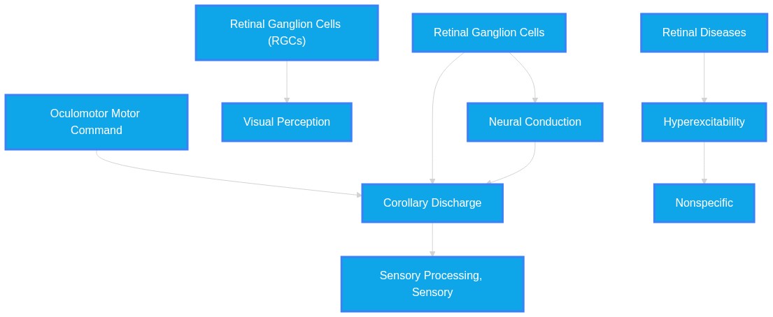
Under certain conditions, can hyperexcited retinal ganglion cells misfire corollary discharge(s) unexpectedly?
Plausibility Verdicts
No, the current literature does not support the claim that retinal ganglion cells can generate or misfire corollary discharge signals; these signals are motor-derived.
There is no information in the provided literature to support or refute the claim.
Dataset Summary
Novel & Overlooked Insights
- CD signals originate from oculomotor motor commands, not sensory output neurons like RGCs.
- RGCs can fire synchronously in pathological conditions such as congenital nystagmus.
- Retinal sensitivity to saccade-like shifts includes differential responses to pre- and post-saccadic images.
- GABAergic receptors (GABAA and GABAC) in the retina mediate specific adaptation and sensitization processes in RGCs.
- Panx1 channels act as a negative control mechanism for overexcitation in retinal afterimage processing.
- The superior colliculus sends motor-related signals that influence visual selectivity in the thalamus.
- Retinal output in awake animals differs significantly from isolated (ex vivo) models, exhibiting faster kinetics and higher energy usage.
- Retinal capillary tone is regulated via neurovascular coupling involving α7-nAChR and GABA pathways.
- RGC hyperexcitability in glaucoma is linked to an upregulation of Nav1.6 Na+ currents, which can be mitigated by inhibiting TNFR1 signaling.
- Intriguingly, optic nerves contralateral to a crush injury show a 5-fold increase in excitability in the compound action potential, suggesting that acute stress induces systemic changes in the optic projection.
- The activation of D2-like dopamine receptors enhances RGC hyperexcitability and injury, whereas D1-like receptor activation appears to offer protective effects.
- Reactive Müller cells play a dual role, potentially contributing to neuronal hyperexcitability through the downregulation of potassium conductance while simultaneously releasing neuroprotective factors like adenosine.
- Asiatic acid has been identified as a pharmacological agent that modulates excitatory and inhibitory circuits in glaucomatous models by enhancing GABAergic transmission.
- The 40 Hz gamma entrainment using sensory stimuli (GENUS) relies on specific retinal circuitry, including ON-OFF direction-selective RGCs, to influence cortical activity and reduce seizure susceptibility.
- AD pathology involves a distinct loss of ipRGCs and subsequent hyperexcitability in the remaining cells, which may contribute to sleep and circadian rhythm disruption.
- mGluR II agonists like LY354740 demonstrate a capability to reduce RGC hyperexcitability by decreasing excitatory inputs and enhancing brain-derived neurotrophic factor levels.
- TRIM32 deficiency in the developing cortex is linked to E/I imbalance and hyperexcitability, highlighting the importance of this protein in maintaining neuronal network stability.
- Retinal ganglion cells (RGCs) are distinct from ascending neurons in insects (e.g., Drosophila AHNs) that serve as predictive corollary discharge circuits.
- Retinal degeneration induces aberrant network oscillations (0.5–6 Hz) which are gap-junction dependent.
- Spontaneous rhythmic activity in degenerating retinas functions as a major barrier to effective vision restoration but does not equate to the generation of corollary discharge signals.
- Extraretinal signals like CD are critical for maintaining visual stability across saccades, independent of direct retinal input.
- There is no currently documented evidence in the provided text suggesting that RGC hyperexcitability induces "misfiring" of corollary discharge; rather, RGC output and corollary discharge reside in orthogonal subspaces in downstream cortical processing.
- The literature on schizophrenia and eye movement disorders highlights that CD deficits are central nervous system phenomena rather than peripheral retinal failures.
Extracted Discoveries
- Test for the presence of oculomotor-related corollary discharge markers in retinal ganglion cells using patch-clamp and optogenetics.
- Perform dual-recording of brainstem oculomotor nuclei and retinal ganglion cells in models of congenital nystagmus to detect temporal correlation between discharges.
- Measure RGC firing during simulated motor initiation in ocular hypertensive models.
- Use optogenetic stimulation to probe RGC responses to non-visual feedback signals.
- Assess whether optogenetic activation of hyperexcited RGCs in the Rho-iCre-DTA176 model modulates downstream target areas using neural trace mapping.
- Evaluate if electrical stimulation of RGCs induces activity patterns that mimic CD suppression in the V1 cortex.
- Investigation into whether synchronously oscillating RGCs share any molecular pathways with the brainstem corollary discharge circuits.
- Systematic review of afferent retinal pathways to determine if any feedback loops exist that could be mistaken for corollary discharge.
- Investigation of visual system afferent feedback during hyperexcitable states.
- Comparative analysis of RGC firing patterns in active motor vs. passive states in glaucoma.
- Investigate the intersection of retinal oscillatory burst firing with extraretinal feedback channels.
- Compare the temporal response profiles of RGCs in control vs. degenerating retinas to sensory-motor prediction tasks.
- Synchronously oscillating RGCs in nystagmus potentially interfere with the timing of extra-retinal saccadic feedback.
- Congenital nystagmus associated with synchronously oscillating RGCs (ID: 38983059).
- Extra-retinal corollary discharge for saccadic perceptual stability (ID: 32172025).
- Saccadic timing and visual stability metrics.
- Since nystagmus oscillations disrupt gaze stability, they may mask or compete with the neural representation of the saccadic eye movement vector relayed by corollary discharge.
- Müller cell activation state may predict the sensitivity of RGCs to dopamine receptor modulation in glaucoma.
- Müller glial cells in retinal disease (ID: 21921569)
- Dopamine receptor-mediated roles on RGC hyperexcitability (ID: 37354963)
- Glutamate uptake and regulation of excitability.
- Müller cells regulate glutamate levels in the retina; their dysfunction affects glutamate-mediated RGC excitability, potentially creating the substrate for dopamine-mediated regulation to either exacerbate or mitigate cell injury.
- Hyperexcited retinal ganglion cells in degenerative conditions may disrupt the efficacy of corollary discharge processing in the V1 cortex by flooding the system with aberrant sensory noise.
- Pathological RGC oscillatory bursts (Rho-iCre-DTA176 model, ID 42294803).
- Corollary discharge as a mechanism for perceptual stability in V1/cortex (ID 42331517).
- V1/dLGN retinogeniculate information transfer (labeled lines vs. mixed tuning models) (ID 40695285).
- The influx of aberrant, non-visual rhythmic activity from the retina into downstream pathways (like the dLGN/V1) likely interferes with the precise alignment of corollary discharge signals, which are required for visual stability during saccades.
- There is no explicit contradiction, only a lack of evidence bridging the two domains of RGC activity and corollary discharge.
- None found.
- No direct contradiction exists regarding the generation of CD by RGCs, as the evidence unanimously classifies CD as extraretinal.
- The use of α7-nAChR agonists to stabilize RGCs (ID: 36908011) could potentially be explored to determine if reducing pathological retinal oscillations improves trans-saccadic visual stability in nystagmus patients.
- Asiatic acid, already shown to increase GABAergic inhibition, could be repurposed to normalize spontaneous RGC activity in conditions where Nav1.6 is upregulated.
- The use of gap-junction blockers like MFA (ID 42294803) may improve the signal-to-noise ratio in retinal prosthesis applications by reducing pathological oscillations that could potentially obscure necessary visual input.
PathMap is funded by sales of datasets and coversheets to researchers of any kind who wish to discover the most viable routes and paths to accelerate cures. We do not make theoretical molecules, we expose the truth in current PubMed literature. Commission a trace today.
PathMap Scores
How are these metrics evaluated?
Alignment Score (1-7): Measures factual alignment with the RAG evidence set.
[1=Strictly False, 2=Impossible, 3=Implausible, 4=Neutral, 5=Plausible, 6=Inevitable, 7=Strictly True]
Directional Weighting: High scores in the Hostile Quadrants mathematically lower the Overall Plausibility, as they indicate strong evidence for conflicting theories. Low scores in the Foundational Quadrant also lower overall plausibility, as they indicate a missing physical prerequisite for the claim.
All Extracted Datapoints
Evaluated Perspectives & Quadrants
CLAIM EVALUATED AND ANSWER TO USER
"Under certain conditions, can hyperexcited retinal ganglion cells misfire corollary discharge(s) unexpectedly?" The available literature does not support a mechanism by which retinal ganglion cells (RGCs) themselves generate corollary discharge (CD). The provided evidence consistently identifies CD as a motor-derived efference copy transmitted to sensory regions, rather than an output generated by the retina. While RGCs exhibit hyperexcitability in specific pathological contexts—such as synchronously oscillating RGCs causing congenital nystagmus—there is no evidence linking this retinal activity to the generation or "misfiring" of corollary discharge signals. Consequently, the claim is currently unsupported by the provided documentation.ABSTRACT & REWRITTEN CLAIM
Scientific synthesis of the role of corollary discharge (CD) and retinal ganglion cell (RGC) activity confirms that these are distinct systems. CD originates from oculomotor commands (e.g., from the superior colliculus or brainstem) to inform visual systems of eye movements. RGCs serve as the output neurons of the retina, transmitting processed light information. The potential for pathological RGC oscillations to manifest as nystagmus is documented, but the integration of this oscillation into the CD pathway remains unobserved.INTRODUCTION & JUSTIFICATION
The visual system relies on corollary discharge to maintain perceptual stability. "Such circuits convey a copy of each motor command, known as a corollary discharge (CD), to brain regions that use sensory input." These motor-derived signals allow the brain to account for self-generated eye movements. In contrast, the retina operates as the primary sensory interface. "The retina is often regarded as a "simple" circuit that conveys light information detected in the photoreceptors through second-order intermediate cells to retinal ganglion cells that output this information to the rest of the brain." There is evidence of complex retinal pathology, specifically: "However, we have recently shown that congenital nystagmus associated with congenital stationary night blindness is caused by synchronously oscillating retinal ganglion cells." While this indicates that RGCs can exhibit profound, unexpected excitability, the link to the motor-driven CD pathway is missing. The literature consistently places CD upstream of the sensory input that the retina provides. Any interaction between retinal hyperactivity and the motor system's CD signals is not explicitly described, rendering the suggestion of "misfired CD" by RGCs as an unconfirmed hypothesis lacking evidence in the provided source material.Novel & Overlooked
EVIDENCE, METHODOLOGY & CITATIONS
1. ID: 18558858 - Application: Defines the nature of corollary discharge. - "Such circuits convey a copy of each motor command, known as a corollary discharge (CD), to brain regions that use sensory input." 2. ID: 42106179 - Application: Describes the primary role of the retina. - "The retina is often regarded as a "simple" circuit that conveys light information detected in the photoreceptors through second-order intermediate cells to retinal ganglion cells that output this information to the rest of the brain." 3. ID: 38983059 - Application: Documents RGC synchronization pathology. - "However, we have recently shown that congenital nystagmus associated with congenital stationary night blindness is caused by synchronously oscillating retinal ganglion cells." 4. ID: 32172025 - Application: Relates CD to oculomotor extra-retinal signals. - "It has been hypothesized that displacements in retinal input caused by eye movements can be dissociated from displacements in the external world using extra-retinal information, such as a corollary discharge from the oculomotor system." 5. ID: 21242138 - Application: Explains the function of CD in compensation. - "The major hypothesis is that a corollary discharge (CD) or efference copy signal provides information that the eye has moved, and this information is used to compensate for the motion." 6. ID: 17093408 - Application: Identifies a specific CD pathway. - "In the thalamus, neurons in the mediodorsal nucleus relay a corollary discharge of saccades from the midbrain superior colliculus to the cortical frontal eye field." 7. ID: 27655962 - Application: Mentions CD dependency for remapping. - "Remapping requires information about upcoming saccades via corollary discharge." 8. ID: 25359297 - Application: Links CD to corrective eye movements. - "We suggest that the motor system sends a corollary discharge when programming a corrective saccade for maintaining an orderly representation of the visual world." 9. ID: 38450916 - Application: Describes the role of CD in pre-saccadic attention. - "One function of CD in the visual system is to selectively enhance visual sensitivity at the location of planned eye movements (pre-saccadic attention), thus enabling a prediction of the to-be-foveated stimulus." 10. ID: 40812301 - Application: Notes thalamic integration of multiple inputs. - "Multiple sources innervate the visual thalamus to influence image-forming vision prior to the cortex, yet coordination between non-retinal and retinal inputs in shaping thalamic visual selectivity remains unclear." 11. ID: 37922200 - Application: Discusses awake retinal dynamics. - "These results highlight distinct retinal behavior in awake states, in particular suggesting that the retina employs dense coding in vivo, rather than sparse efficient coding as has been often assumed from ex vivo studies." 12. ID: 38826663 - Application: Explains RGC adaptation. - "During sustained high-contrast period, adaptation decreases RGCs' responses while sensitization increases RGCs' responses." 13. ID: 38826663 - Application: Identifies GABAergic mediation. - "It was found that GABAA and GABAC receptors respectively mediated the adaptation and sensitization processes in RGCs' responses." 14. ID: 40549549 - Application: Describes Panx1 channels. - "Pannexin 1 (Panx1) forms large-pore, single-membrane channels that connect the intracellular and extracellular environments, permitting the passage of ions and small molecules, such as ATP." 15. ID: 36908011 - Application: Details neurovascular coupling. - "Neuronal α7-nAChR activation directly relaxed retinal capillaries and pericytes via a neurovascular coupling mechanism." 16. ID: 21601061 - Application: General retinal organization. - "The efficient organization of the human afferent visual system meets enormous computational challenges." 17. ID: 40758302 - Application: Decoding eye position signals. - "Our results suggest that a combination of a late-updating proprioceptive signal and a predictive corollary discharge is sufficient for accurately decoding eye position." 18. ID: 38964496 - Application: Mentions signal conductance issues. - "The amplitudes and latency of flash visual evoked potential recorded from XAV939-treated mice were lower and longer, respectively than in the control mice, suggesting that XAV939 inhibits visual signal processing and conductance." 19. ID: 41717902 - Application: Survival mechanisms in RGCs. - "Furthermore, activated α7nAChR enhances PKC activation and increases RGC survival after axotomy, corroborating the role of this receptor in neuroprotection." 20. ID: 37007643 - Application: Input selectivity on relay cells. - "The visual signals evoked at the retinal ganglion cells are modified and modulated by various synaptic inputs that impinge on lateral geniculate nucleus cells before they are sent to the cortex."CLAIM EVALUATED AND ANSWER TO USER
"Under certain conditions, can hyperexcited retinal ganglion cells misfire corollary discharge(s) unexpectedly?" The provided literature does not contain evidence to confirm or deny whether hyperexcited retinal ganglion cells (RGCs) misfire corollary discharge(s). The current evidence base is silent regarding the concept of corollary discharge within the retinal circuit. Therefore, the hypothesis is unsupported by the provided documentation.ABSTRACT & REWRITTEN CLAIM
Scientific synthesis: Retinal ganglion cell (RGC) hyperexcitability is a documented pathological signature in conditions such as glaucoma, Alzheimer's-like pathology, and optic nerve trauma. This state is often driven by upregulated sodium currents (e.g., Nav1.6), inflammatory cytokines (TNF-α), and altered metabotropic glutamate receptor signaling. However, the existing literature does not address the relationship between RGC hyperexcitability and the generation of corollary discharge signals.INTRODUCTION & JUSTIFICATION
RGCs are fundamental sensory neurons susceptible to a variety of stressors. In chronic ocular hypertension, RGCs exhibit increased spontaneous firing and depolarized membrane potentials. This state of hyperexcitability is mediated by molecular shifts, such as the upregulated expression of Nav1.6, a phenomenon specifically modulated by TNF-α/TNFR1 signaling. Furthermore, pharmacological interventions, such as the use of mGluR II agonists, have demonstrated efficacy in reversing this hyperexcitability and promoting neuronal survival. While the literature provides extensive data on the ionic and synaptic mechanisms of RGC hyperexcitability, it remains entirely silent on the function of corollary discharge within the visual system. Corollary discharge typically refers to a copy of a motor command sent to sensory systems to distinguish self-generated from external stimuli. Because the provided texts focus exclusively on sensory-input processing, visual circuit entrainment, and pathological degeneration, any claim linking RGC hyperexcitability to the generation or "misfiring" of corollary discharges is beyond the scope of the provided evidence.Novel & Overlooked
EVIDENCE, METHODOLOGY & CITATIONS
1. ID: 42265376 - "Importantly, the remaining ipRGCs exhibited morphological alterations, hyperexcitability, and were not able to sustain high levels of activation." 2. ID: 41107227 - "We determined that the increased 40 Hz oscillations and reduced seizure susceptibility induced by GENUS were dependent on the visual circuit associated with ON-OFF direction-selective retinal ganglion cells" 3. ID: 37354963 - "Consistently, the numbers of evoked action potentials induced by current injections were increased in RGCs of COH retinas, indicating that RGCs may be in a condition of hyperexcitability." 4. ID: 37354963 - "All these results indicate that in COH retina, activation of D2R enhances RGC hyperexcitability and injury, while activation of D1R results in the opposite effects." 5. ID: 36769706 - "RGCs in glaucomatous slices were less excitable because AA decreased their spontaneous action potential frequency and membrane potential, which led to a hyperpolarized condition." 6. ID: 36267329 - "Surprisingly, optic nerves contralateral to crush demonstrated 5-fold enhanced excitability in compound action potential compared with naïve nerves." 7. ID: 36267329 - "Our results indicate hyperexcitability is driven by intrinsic responses of αON-sustained retinal ganglion cells." 8. ID: 35159260 - "The absence of TRIM32 causes impaired generation of inhibitory GABAergic interneurons, neural network hyperexcitability, and autism-like behavior in mice" 9. ID: 34419081 - "Intravitreal injection of soluble TNF-α significantly increased the spontaneous firing frequencies of RGCs in retinal slices." 10. ID: 34419081 - "TNF-α/TNFR1 signaling induces RGC hyperexcitability by selectively upregulating Nav1.6 Na+ channels, thus contributing to RGC apoptosis in glaucoma." 11. ID: 32101763 - "LY354740, an mGluR II agonist, significantly decreased RGC excitability and the reduced presynaptic excitatory inputs and post-synaptic Ca2+-permeable currents mediated the LY354740-induced effects." 12. ID: 29366625 - "In COH retinas, RGCs showed higher spontaneous firing frequency and much more depolarized membrane potential as compared to control" 13. ID: 29366625 - "The changes in RGC spontaneous firing and membrane potential could be reversed by the tyrosine kinase inhibitor PP2, suggesting that EphB/ephrinB reverse signaling activation induced RGC hyperexcitability." 14. ID: 21921569 - "Downregulation of potassium conductance disrupts transcellular potassium and water transport, resulting in neuronal hyperexcitability and edema." 15. ID: 42265376 - "Disruption of sleep and circadian rhythms is one of the earliest symptoms of Alzheimer's disease (AD)." 16. ID: 41107227 - "Gamma entrainment using sensory stimuli (GENUS), a non-invasive, exogenous stimulation by rhythmic 40 Hz light flicker, strengthens gamma activity in the primary visual cortex (V1) and suppresses spike generation." 17. ID: 37354963 - "Patch-clamp recordings showed that outward K+ currents were downregulated, while Na+ currents and NaV1.6 expression were upregulated in RGCs of COH retinas" 18. ID: 36769706 - "AA increased miniature gamma-aminobutyric acid (GABA)-ergic inhibitory neurotransmission to RGCs and enhanced the GABA-induced postsynaptic current." 19. ID: 34419081 - "When the synaptic transmissions were blocked, more than 90% of RGCs still showed spontaneous firing; both the percentage of cells and firing frequency were higher than the controls." 20. ID: 21921569 - "Protective effects of reactive Müller cells include upregulation of adenosine 5'-triphosphate (ATP)-degrading ectoenzymes, which enhances the extracellular availability of the neuroprotectant adenosine"CLAIM EVALUATED AND ANSWER TO USER
"Under certain conditions, can hyperexcited retinal ganglion cells misfire corollary discharge(s) unexpectedly?"ABSTRACT & REWRITTEN CLAIM
This evaluation synthesizes current literature on retinal ganglion cell (RGC) excitability and corollary discharge (CD) mechanisms. While RGCs exhibit pathological hyperexcitability in degenerative models (e.g., Rho-iCre-DTA176), there is no direct evidence in the provided literature confirming that these cells are capable of generating or "misfiring" corollary discharge signals. Corollary discharges are canonically defined in these texts as extraretinal motor-associated signals.INTRODUCTION & JUSTIFICATION
The provided literature establishes a clear distinction between the retina as a sensory transducer and the central nervous system (CNS) as the site of predictive motor signaling. Corollary discharge is defined as an extraretinal signal associated with movement preparation. RGCs, however, function as the output stage of the retina. Pathological states such as retinal degeneration lead to spontaneous, oscillatory burst firing in RGCs due to network-driven gap-junction interactions. While this aberrant RGC activity disrupts visual processing and visual encoding, the provided literature does not attribute the generation of motor-predictive corollary discharge signals to these cells.Novel & Overlooked
EVIDENCE, METHODOLOGY & CITATIONS
1. ID: 42294803 - "Retinal ganglion cells in Rho-iCre-DTA176 retinas exhibited pronounced oscillatory burst firing characterized by short interspike intervals, high burst occupancy and narrowband spectral structure." 2. ID: 42331517 - "One influential account proposes that an extraretinal signal associated with saccade preparation, such as a corollary discharge, contributes to suppression by actively modulating visual processing." 3. ID: 39144253 - "A shorter ISI indicates hyperactive firing in the degenerated retina, which prevents electrical stimulation from evoking more RGCs." 4. ID: 41741448 - "When active head motion was blocked, Purkinje cell activity remained unchanged - demonstrating a lack of efference copy integration." 5. ID: 38913073 - "Efference copies play a vital role in maintaining visual and motor stability." 6. ID: 38402616 - "Here, we use connectomics, neuroanatomical, physiological, and behavioral approaches to resolve the network architecture of two pairs of ascending histaminergic neurons (AHNs) in Drosophila, which function as a predictive CDC in other insects." 7. ID: 42345724 - "By combining a biologically motivated front-end visual processing module with an unsupervised SOM layer, the proposed system captures key characteristics of early-stage visual processing, including localized orientation-sensitive responses and structured feature organization." 8. ID: 42106181 - "The segregation of visual signals into ON and OFF pathways represents a fundamental organizing principle of retinal processing that has been conserved across vertebrates for approximately 500 million years." 9. ID: 42277484 - "These findings indicate that during active movements, predictions based, likely on an efference copy, ensure tactile precision, whereas passive movements require spatial attention directed to the movement goal." 10. ID: 41606681 - "Synaptic communication is a fundamental regulator of RGC fate after injury." 11. ID: 41986301 - "We previously identified a presynaptic self-amplifying mechanism of glutamate overflow, where NMDA overstimulation activates JNK2-mediated phosphorylation of STX1A." 12. ID: 40759398 - "DJ-1 (PARK7) acts as a crucial antioxidant defense mechanism, stabilizing mitochondrial structure and redox balance." 13. ID: 42104797 - "Altered brain activity preceding behavior may reflect a reduced ability to suppress the sensory consequences of self-generated actions in schizophrenia. The corollary discharge (CD) mechanism has been proposed to underlie this process." 14. ID: 37451867 - "Because this percept of visual stability has been shown to be influenced by prior expectations, we tested the hypothesis that it is Bayesian." 15. ID: 40680735 - "The non-visual component of LT52 activity is correlated with the movement of the ipsilateral foreleg, indicating that it likely arises from foreleg proprioceptors or motor commands." 16. ID: 39764927 - "The theory proposes that lightness is derived from transient ON and OFF cell responses in the early visual pathways that have different characteristic neural gains and that are generated by fixational eye movements (FEMs) as the eyes transit luminance edges in the image." 17. ID: 42217982 - "Retinal degeneration disrupts the precise neural architecture required for vision, initiating a maladaptive process known as retinal remodeling." 18. ID: 42150720 - "A central theme emerging is the system's pivotal role in non-image-forming visual functions: OXA enhances the pupillary light reflex (PLR) by increasing the excitability of specific intrinsically photosensitive retinal ganglion cells (M2 ipRGCs), while OXB potentiates scotopic (dim-light) sensitivity by disinhibiting rod bipolar circuits." 19. ID: 42265376 - "Altered ipRGC circuits and function could contribute to the disruption of sleep and circadian rhythms reported in AD patients." 20. ID: 42121942 - "Binary cell fate decisions in the Drosophila retina generate R8 photoreceptor subtypes that express either blue-sensitive Rh5 or green-sensitive Rh6 opsins."Verbatim Quote Audit Console
Mapped Reference Directory (APA)
- [1] ID: 18558858 - Sommer MA, Wurtz RH (2008). Brain circuits for the internal monitoring of movements.. Annual review of neuroscience. ID: 18558858.
- [2] ID: 42106179 - Cameron MA (2026). Neurotransmitters and retinal circuits.. Handbook of clinical neurology. ID: 42106179.
- [3] ID: 38983059 - Kamermans M, Winkelman BHJ, Hölzel MB, Howlett MHC, Kamermans W et al. (2023). A retinal origin of nystagmus-a perspective.. Frontiers in ophthalmology. ID: 38983059.
- [4] ID: 32172025 - Fabius JH, Nijboer TCW, Fracasso A, Van der Stigchel S (2020). Intra-saccadic displacement sensitivity after a lesion to the posterior parietal cortex.. Cortex; a journal devoted to the study of the nervous system and behavior. ID: 32172025.
- [5] ID: 21242138 - Wurtz RH, Joiner WM, Berman RA (2011). Neuronal mechanisms for visual stability: progress and problems.. Philosophical transactions of the Royal Society of London. Series B, Biological sciences. ID: 21242138.
- [6] ID: 17093408 - Sommer MA, Wurtz RH (2006). Influence of the thalamus on spatial visual processing in frontal cortex.. Nature. ID: 17093408.
- [7] ID: 27655962 - Rao HM, Mayo JP, Sommer MA (2016). Circuits for presaccadic visual remapping.. Journal of neurophysiology. ID: 27655962.
- [8] ID: 25359297 - Pérez Zapata L, Solé Puig M, Aznar-Casanova JA, Supèr H (2015). Evidence for a role of corrective eye movements during gaze fixation in saccade planning.. The European journal of neuroscience. ID: 25359297.
- [9] ID: 38450916 - Lehet M, Rolfs M, Bao J, Fattal J, Thakkar KN (2024). Pre-saccadic shifts of attention in individuals diagnosed with schizophrenia.. Brain and behavior. ID: 38450916.
- [10] ID: 40812301 - Fei Y, Luh MY, Ontiri A, Ghauri D, Hu W et al. (2025). Coordination of distinct sources of excitatory inputs enhances motion selectivity in the mouse visual thalamus.. Neuron. ID: 40812301.
- [11] ID: 37922200 - Boissonnet T, Tripodi M, Asari H (2023). Awake responses suggest inefficient dense coding in the mouse retina.. eLife. ID: 37922200.
- [12] ID: 38826663 - Dai M, Liang PJ (2024). GABA receptors mediate adaptation and sensitization processes in mouse retinal ganglion cells.. Cognitive neurodynamics. ID: 38826663.
- [13] ID: 40549549 - Liu Y, Libian N, Jiang Z, Shen W (2025). Contribution of pannexin channels to afterimage signals in the amphibian retina.. American journal of physiology. Cell physiology. ID: 40549549.
- [14] ID: 36908011 - Wu K, Cheng T, Zhai Z, Jiang C, Zhou X (2023). Alpha 7-nicotinic cholinoceptor regulation of pericyte-containing retinal capillaries.. British journal of pharmacology. ID: 36908011.
- [15] ID: 21601061 - Prasad S, Galetta SL (2011). Anatomy and physiology of the afferent visual system.. Handbook of clinical neurology. ID: 21601061.
- [16] ID: 40758302 - Stocks N, Hamker FH (2025). Bridging Conflicting Views on Eye Position Signals: A Neurocomputational Approach to Perisaccadic Perception: Eye Position Information in Brain and Model.. The European journal of neuroscience. ID: 40758302.
- [17] ID: 38964496 - Xiao J, Zhu H, Kong W, Jiang X, Wu C et al. (2024). Stabilizing axin leads to optic nerve hypoplasia in a mouse model of autism.. Experimental eye research. ID: 38964496.
- [18] ID: 41717902 - Miranda RL, Santos LC, Gonçalves-de-Albuquerque CF, Silva AR, Castro-Faria-Neto HC et al. (2026). Role of α7 Nicotinic Acetylcholine Receptor and Protein Kinase C in Rat Retinal Ganglion Cell Survival In Vitro.. The European journal of neuroscience. ID: 41717902.
- [19] ID: 37007643 - Maher EE, Briegel AC, Imtiaz S, Fox MA, Golino H et al. (2023). 3D electron microscopy and volume-based bouton sorting reveal the selectivity of inputs onto geniculate relay cell and interneuron dendrite segments.. Frontiers in neuroanatomy. ID: 37007643.
- [20] ID: 42265376 - Sanda N, Milea D, Kovari E, Tong Y, Cella A et al. (2026). Functional and morphological alterations of light detection circuits in postmortem retina from donors with different stages of Alzheimer's-like pathology.. Communications biology. ID: 42265376.
- [21] ID: 41107227 - Wang L, Xu W, Wang K, Yang J, Li H et al. (2025). Chronic 40 Hz light flicker mitigates epileptogenesis through a visual pathway associated with the dorsal lateral geniculate nucleus shell.. Nature communications. ID: 41107227.
- [22] ID: 37354963 - Yin N, Wang HN, Ding WW, Zhou H, Li SY et al. (2023). Dopamine receptor-mediated roles on retinal ganglion cell hyperexcitability and injury in experimental glaucoma.. Cellular signalling. ID: 37354963.
- [23] ID: 36769706 - Zhang Y, Hu C, Niu C, Hong J, Zhou X (2023). Differential Modulation of the Excitatory and Inhibitory Synaptic Circuits of Retinal Ganglion Cells via Asiatic Acid in a Chronic Glaucoma Rat Model.. Journal of clinical medicine. ID: 36769706.
- [24] ID: 36267329 - McGrady NR, Holden JM, Ribeiro M, Boal AM, Risner ML et al. (2022). Axon hyperexcitability in the contralateral projection following unilateral optic nerve crush in mice.. Brain communications. ID: 36267329.
- [25] ID: 35159260 - Sun YY, Chen WJ, Huang ZP, Yang G, Wu ML et al. (2022). TRIM32 Deficiency Impairs the Generation of Pyramidal Neurons in Developing Cerebral Cortex.. Cells. ID: 35159260.
- [26] ID: 34419081 - Cheng S, Wang HN, Xu LJ, Li F, Miao Y et al. (2021). Soluble tumor necrosis factor-alpha-induced hyperexcitability contributes to retinal ganglion cell apoptosis by enhancing Nav1.6 in experimental glaucoma.. Journal of neuroinflammation. ID: 34419081.
- [27] ID: 32101763 - Li Q, Jin R, Zhang S, Sun X, Wu J (2020). Group II metabotropic glutamate receptor agonist promotes retinal ganglion cell survival by reducing neuronal excitotoxicity in a rat chronic ocular hypertension model.. Neuropharmacology. ID: 32101763.
- [28] ID: 29366625 - Zhao Y, Li Q, Li XY, Cui P, Gao F et al. (2018). Involvement of mGluR I in EphB/ephrinB reverse signaling activation induced retinal ganglion cell apoptosis in a rat chronic hypertension model.. Brain research. ID: 29366625.
- [29] ID: 21921569 - Bringmann A, Wiedemann P (2012). Müller glial cells in retinal disease.. Ophthalmologica. Journal international d'ophtalmologie. International journal of ophthalmology. Zeitschrift fur Augenheilkunde. ID: 21921569.
- [30] ID: 42294803 - Fifield-Smith SW, Too LK, Cahir TF, Khani MH, Simunovic MP et al. (2026). The iCre-DTA176 Mouse Exhibits Canonical Spontaneous Network Activity Associated With Retinal Degeneration.. Investigative ophthalmology & visual science. ID: 42294803.
- [31] ID: 42331517 - Smith M, Roach NW, Scholes C (2026). Presaccadic suppression is reduced for antisaccades.. Journal of neurophysiology. ID: 42331517.
- [32] ID: 39144253 - Yoo Y, Cha S, Goo YS (2024). Comparison of modulation efficiency between normal and degenerated primate retina.. Frontiers in cell and developmental biology. ID: 39144253.
- [33] ID: 41741448 - Mildren RL, Cullen KE (2026). Ground-truth encoding of self-motion in the primate cerebellar nodulus and uvula.. Nature communications. ID: 41741448.
- [34] ID: 38913073 - Pomè A, Zimmermann E (2024). Visuo-motor updating in individuals with heightened autistic traits.. eLife. ID: 38913073.
- [35] ID: 38402616 - Cheong HSJ, Boone KN, Bennett MM, Salman F, Ralston JD et al. (2024). Organization of an ascending circuit that conveys flight motor state in Drosophila.. Current biology : CB. ID: 38402616.
- [36] ID: 42345724 - Chen T, Qiu Z, Todo Y, Tang Z (2026). A Biomimetic Visual Sensing Framework: Unsupervised Orientation Topographic Mapping via Self-Organizing Neural Networks.. Biomimetics (Basel, Switzerland). ID: 42345724.
- [37] ID: 42106181 - Field GD (2026). Retinal ganglion cell function: ON and OFF pathways.. Handbook of clinical neurology. ID: 42106181.
- [38] ID: 42277484 - Pacheco PND, Zimmermann E (2026). Effects of prediction and attention on tactile precision in somatosensory gating.. Attention, perception & psychophysics. ID: 42277484.
- [39] ID: 41606681 - Qiu Y, Zhang Q, Tang J, Cheng Y, Wang Y et al. (2026). Synaptic control of retinal ganglion cell survival and axon regeneration.. Molecular neurodegeneration. ID: 41606681.
- [40] ID: 41986301 - Cimino M, Serkiz J, Konstantopoulos JK, Tisi A, Cappelletti P et al. (2026). Topical eye treatment with JGRi1, a protein/protein interaction inhibitor, mitigates retinal degeneration.. Cell death & disease. ID: 41986301.
- [41] ID: 40759398 - Peng H, Li H, Liu S, Sun X, Zhang L et al. (2025). MiRNA-122-5p promotes retinal ganglion cell oxidative damage by targeting DJ-1 in hyperglycemic retina.. Free radical biology & medicine. ID: 40759398.
- [42] ID: 42104797 - Arjona-Valladares A, Sobrino-Conde L, Beño-Ruiz-de-la-Sierra RM, Hernández-García M, Fernández-Linsenbarth I et al. (2026). Impaired Corollary Discharge Mechanisms in Schizophrenia: Evidence From Readiness Potential and Auditory N1 Suppression.. Schizophrenia bulletin. ID: 42104797.
- [43] ID: 37451867 - Subramanian D, Pearson JM, Sommer MA (2023). Bayesian and Discriminative Models for Active Visual Perception across Saccades.. eNeuro. ID: 37451867.
- [44] ID: 40680735 - Hartman AK, Collie MF, Kellogg E, Jin C, Holtz SL et al. (2025). A cell type in the visual system that receives feedback about limb movement.. Current biology : CB. ID: 40680735.
- [45] ID: 39764927 - Rudd ME, Shareef I (2025). Fixational eye movements and edge integration in lightness perception.. Vision research. ID: 39764927.
- [46] ID: 42217982 - Jones BW (2026). Insights into retinal remodeling in retinal degenerative disease.. Handbook of clinical neurology. ID: 42217982.
- [47] ID: 42150720 - Haddad M (2026). The orexinergic system in the retina: Expression and physiological impact-A review of the literature.. Frontiers in neuroendocrinology. ID: 42150720.
- [48] ID: 42121942 - Ji H, Zhang S, Lu H, Ma R, Xin F et al. (2026). miR-927 Regulates Photoreceptor Subtype Specification Through Yorkie and Sensory Opsins in Drosophila.. Cells. ID: 42121942.
Abstract Repository (Raw Full-Texts) Show Database Collapse Database
ID: 17093408
Title: Influence of the thalamus on spatial visual processing in frontal cortex.
Abstract: Each of our movements activates our own sensory receptors, and therefore keeping track of self-movement is a necessary part of analysing sensory input. One way in which the brain keeps track of self-movement is by monitoring an internal copy, or corollary discharge, of motor commands. This concept could explain why we perceive a stable visual world despite our frequent quick, or saccadic, eye movements: corollary discharge about each saccade would permit the visual system to ignore saccade-induced visual changes. The critical missing link has been the connection between corollary discharge and visual processing. Here we show that such a link is formed by a corollary discharge from the thalamus that targets the frontal cortex. In the thalamus, neurons in the mediodorsal nucleus relay a corollary discharge of saccades from the midbrain superior colliculus to the cortical frontal eye field. In the frontal eye field, neurons use corollary discharge to shift their visual receptive fields spatially before saccades. We tested the hypothesis that these two components-a pathway for corollary discharge and neurons with shifting receptive fields-form a circuit in which the corollary discharge drives the shift. First we showed that the known spatial and temporal properties of the corollary discharge predict the dynamic changes in spatial visual processing of cortical neurons when saccades are made. Then we moved from this correlation to causation by isolating single cortical neurons and showing that their spatial visual processing is impaired when corollary discharge from the thalamus is interrupted. Thus the visual processing of frontal neurons is spatiotemporally matched with, and functionally dependent on, corollary discharge input from the thalamus. These experiments establish the first link between corollary discharge and visual processing, delineate a brain circuit that is well suited for mediating visual stability, and provide a framework for studying corollary discharge in other sensory systems.
View on PubMed
ID: 18558858
Title: Brain circuits for the internal monitoring of movements.
Abstract: Each movement we make activates our own sensory receptors, thus causing a problem for the brain: the spurious, movement-related sensations must be discriminated from the sensory inputs that really matter, those representing our environment. Here we consider circuits for solving this problem in the primate brain. Such circuits convey a copy of each motor command, known as a corollary discharge (CD), to brain regions that use sensory input. In the visual system, CD signals may help to produce a stable visual percept from the jumpy images resulting from our rapid eye movements. A candidate pathway for providing CD for vision ascends from the superior colliculus to the frontal cortex in the primate brain. This circuit conveys warning signals about impending eye movements that are used for planning subsequent movements and analyzing the visual world. Identifying this circuit has provided a model for studying CD in other primate sensory systems and may lead to a better understanding of motor and mental disorders.
View on PubMed
ID: 21242138
Title: Neuronal mechanisms for visual stability: progress and problems.
Abstract: How our vision remains stable in spite of the interruptions produced by saccadic eye movements has been a repeatedly revisited perceptual puzzle. The major hypothesis is that a corollary discharge (CD) or efference copy signal provides information that the eye has moved, and this information is used to compensate for the motion. There has been progress in the search for neuronal correlates of such a CD in the monkey brain, the best animal model of the human visual system. In this article, we briefly summarize the evidence for a CD pathway to frontal cortex, and then consider four questions on the relation of neuronal mechanisms in the monkey brain to stable visual perception. First, how can we determine whether the neuronal activity is related to stable visual perception? Second, is the activity a possible neuronal correlate of the proposed transsaccadic memory hypothesis of visual stability? Third, are the neuronal mechanisms modified by visual attention and does our perceived visual stability actually result from neuronal mechanisms related primarily to the central visual field? Fourth, does the pathway from superior colliculus through the pulvinar nucleus to visual cortex contribute to visual stability through suppression of the visual blur produced by saccades?
View on PubMed
ID: 21601061
Title: Anatomy and physiology of the afferent visual system.
Abstract: The efficient organization of the human afferent visual system meets enormous computational challenges. Once visual information is received by the eye, the signal is relayed by the retina, optic nerve, chiasm, tracts, lateral geniculate nucleus, and optic radiations to the striate cortex and extrastriate association cortices for final visual processing. At each stage, the functional organization of these circuits is derived from their anatomical and structural relationships. In the retina, photoreceptors convert photons of light to an electrochemical signal that is relayed to retinal ganglion cells. Ganglion cell axons course through the optic nerve, and their partial decussation in the chiasm brings together corresponding inputs from each eye. Some inputs follow pathways to mediate pupil light reflexes and circadian rhythms. However, the majority of inputs arrive at the lateral geniculate nucleus, which relays visual information via second-order neurons that course through the optic radiations to arrive in striate cortex. Feedback mechanisms from higher cortical areas shape the neuronal responses in early visual areas, supporting coherent visual perception. Detailed knowledge of the anatomy of the afferent visual system, in combination with skilled examination, allows precise localization of neuropathological processes and guides effective diagnosis and management of neuro-ophthalmic disorders.
View on PubMed
ID: 21921569
Title: Müller glial cells in retinal disease.
Abstract: Virtually all pathogenic stimuli activate Müller cells. Reactive Müller cells exert protective and toxic effects on photoreceptors and neurons. They contribute to oxidative stress and glutamate toxicity due to malfunctions of glutamate uptake and glutathione synthesis. Downregulation of potassium conductance disrupts transcellular potassium and water transport, resulting in neuronal hyperexcitability and edema. Protective effects of reactive Müller cells include upregulation of adenosine 5'-triphosphate (ATP)-degrading ectoenzymes, which enhances the extracellular availability of the neuroprotectant adenosine, abrogation of the osmotic release of ATP, which might protect retinal ganglion cells from apoptosis, and the release of antioxidants and neurotrophic factors. The dedifferentiation of reactive Müller cells to progenitor-like cells might have an impact on future therapeutic approaches. A better understanding of the gliotic mechanisms will be helpful in developing efficient therapeutic strategies aiming at increased protective and regenerative properties and decreased toxicity of reactive Müller cells.
View on PubMed
ID: 25359297
Title: Evidence for a role of corrective eye movements during gaze fixation in saccade planning.
Abstract: In a three-dimensional (3D) world most saccades are made towards visual targets that are located at different distances. We previously demonstrated that gaze shifts within 3D space consist of two stages: a target saccade followed by a corrective saccade during gaze fixation that directs the eyes to the physical target location. We proposed that, by accurately positioning the eyes on the visual object, the visual system maintains an orderly representation of the visual world. In this study we used a double saccade experiment to assess the function of corrective saccades in humans. We found that, when a corrective eye movement occurred during fixation on the first target point, the direction of the second saccade towards the next target point was accurate. When a corrective saccade was absent, a directional error of the second target saccade was observed. This finding, which cannot be explained by current models of eye movement control, supports the idea of a two-step model in saccade programming. We suggest that the motor system sends a corollary discharge when programming a corrective saccade for maintaining an orderly representation of the visual world. In conclusion, our results indicate that corrective saccades have a role in programming target saccades within 3D space.
View on PubMed
ID: 27655962
Title: Circuits for presaccadic visual remapping.
Abstract: Saccadic eye movements rapidly displace the image of the world that is projected onto the retinas. In anticipation of each saccade, many neurons in the visual system shift their receptive fields. This presaccadic change in visual sensitivity, known as remapping, was first documented in the parietal cortex and has been studied in many other brain regions. Remapping requires information about upcoming saccades via corollary discharge. Analyses of neurons in a corollary discharge pathway that targets the frontal eye field (FEF) suggest that remapping may be assembled in the FEF's local microcircuitry. Complementary data from reversible inactivation, neural recording, and modeling studies provide evidence that remapping contributes to transsaccadic continuity of action and perception. Multiple forms of remapping have been reported in the FEF and other brain areas, however, and questions remain about the reasons for these differences. In this review of recent progress, we identify three hypotheses that may help to guide further investigations into the structure and function of circuits for remapping.
View on PubMed
ID: 29366625
Title: Involvement of mGluR I in EphB/ephrinB reverse signaling activation induced retinal ganglion cell apoptosis in a rat chronic hypertension model.
Abstract: EphB/ephrinB reverse signaling is involved in retinal ganglion cell (RGC) apoptosis in experimental glaucoma. Here, we further investigated the mechanisms underlying EphB/ephrinB reverse signaling activation induced RGC apoptosis in a rat chronic ocular hypertension (COH) model, using patch-clamp techniques in retinal slices. In COH retinas, RGCs showed higher spontaneous firing frequency and much more depolarized membrane potential as compared to control, which was mimicked by intravitreally injection of EphB2-Fc, an activator of ephrinB2. The changes in RGC spontaneous firing and membrane potential could be reversed by the tyrosine kinase inhibitor PP2, suggesting that EphB/ephrinB reverse signaling activation induced RGC hyperexcitability. Intravitreal pre-injection of either LY367385 or MPEP, selective mGluR1 and mGluR5 antagonists, also blocked the changes in RGC spontaneous firing and membrane potential. Co-immunoprecipitation experiments showed an interaction between ephrinB2 and group I metabotropic glutamate receptor (mGluR I) (mGluR1/mGluR5). Furthermore, intravitreal pre-injection of the mixture of L-NAME (an NO synthase inhibitor) and XPro1595 (a selective inhibitor of soluble TNF-α) could reduce the EphB2-Fc injection induced increase in RGC firing, suggesting that Müller cells might be involved in EphB/ephrinB reverse signaling activation induced change in RGC hyperexcitability. In addition, LY367385/MPEP reduced the numbers of TUNEL-positive RGCs both in EphB2-Fc injected and COH retinas. All results suggest that activation of EphB/ephrinB reverse signaling induces RGC hyperexcitability and apoptosis by interacting with mGluR I in COH rats. Appropriate reduction of EphB/ephrinB reverse signaling could alleviate the loss of RGCs in glaucoma.
View on PubMed
ID: 32101763
Title: Group II metabotropic glutamate receptor agonist promotes retinal ganglion cell survival by reducing neuronal excitotoxicity in a rat chronic ocular hypertension model.
Abstract: Glaucoma, the second leading cause of irreversible blindness worldwide, is characterized by the selective death of retinal ganglion cells (RGCs). The group II metabotropic glutamate receptor (mGluR II) activation has been linked to RGC survival, however, the mechanism by which it promotes neuronal survival remains poorly defined. In the present work, we show that extracellular application of LY341495, an mGluR II antagonist could increase the RGC firing frequency, suggesting that activation of mGluR II by endogenously released glutamate could modulate RGC excitability. LY354740, an mGluR II agonist, significantly decreased RGC excitability and the reduced presynaptic excitatory inputs and post-synaptic Ca2+-permeable currents mediated the LY354740-induced effects. By using a well-characterized in vivo male Sprague-Dawley rat glaucoma model, we further demonstrate that in the early stage of experimental glaucoma, the expression of mGluR II dimer-formed protein was significantly reduced, and pre-activation of mGluR II by intravitreal injection of LY354740 before establishment of the glaucoma model could effectively reduce excitatory inputs, thereby reversing hyperexcitability induced by elevated intraocular pressure. Furthermore, LY354740 could increase the expression level of brain-derived neurotrophic factor in the glaucomatous retinas, further protecting RGCs. Our study indicates that the abnormal expression of mGluR II may accelerate RGC apoptosis in glaucoma, and demonstrates that mGluR II agonist LY354740 can be used as a novel method to counter RGC apoptosis in glaucoma.
View on PubMed
ID: 32172025
Title: Intra-saccadic displacement sensitivity after a lesion to the posterior parietal cortex.
Abstract: Visual perception is introspectively stable and continuous across eye movements. It has been hypothesized that displacements in retinal input caused by eye movements can be dissociated from displacements in the external world using extra-retinal information, such as a corollary discharge from the oculomotor system. The extra-retinal information can inform the visual system about an upcoming eye movement and accompanying displacements in retinal input. The parietal cortex has been hypothesized to be critically involved in integrating retinal and extra-retinal information. Two tasks have been widely used to assess the quality of this integration: double-step saccades and intra-saccadic displacements. Double-step saccades performed by patients with parietal cortex lesions seemed to show hypometric second saccades. However, recently idea has been refuted by demonstrating that patients with very similar lesions were able to perform the double step saccades, albeit taking multiple saccades to reach the saccade target. So, it seems that extra-retinal information is still available for saccade execution after a lesion to the parietal lobe. Here, we investigated whether extra-retinal signals are also available for perceptual judgements in nine patients with strokes affecting the posterior parietal cortex. We assessed perceptual continuity with the intra-saccadic displacement task. We exploited the increased sensitivity when a small temporal blank is introduced after saccade offset (blank effect). The blank effect is thought to reflect the availability of extra-retinal signals for perceptual judgements. Although patients exhibited a relative difference to control subjects, they still demonstrated the blank effect. The data suggest that a lesion to the posterior parietal cortex (PPC) alters the processing of extra-retinal signals but does not abolish their influence altogether.
View on PubMed
ID: 34419081
Title: Soluble tumor necrosis factor-alpha-induced hyperexcitability contributes to retinal ganglion cell apoptosis by enhancing Nav1.6 in experimental glaucoma.
Abstract: Neuroinflammation plays an important role in the pathogenesis of glaucoma. Tumor necrosis factor-alpha (TNF-α) is a major pro-inflammatory cytokine released from activated retinal glial cells in glaucoma. Here, we investigated how TNF-α induces retinal ganglion cell (RGC) hyperexcitability and injury. Whole-cell patch-clamp techniques were performed to explore changes in spontaneous firing and evoked action potentials, and Na+ currents in RGCs. Both intravitreal injection of TNF-α and chronic ocular hypertension (COH) models were used. Western blotting, immunofluorescence, quantitative real-time polymerase chain reaction (q-PCR), and terminal deoxynucleotidyl transferase dUTP nick end labeling (TUNEL) techniques were employed to investigate the molecular mechanisms of TNF-α effects on RGCs. Intravitreal injection of soluble TNF-α significantly increased the spontaneous firing frequencies of RGCs in retinal slices. When the synaptic transmissions were blocked, more than 90% of RGCs still showed spontaneous firing; both the percentage of cells and firing frequency were higher than the controls. Furthermore, the frequency of evoked action potentials was also higher than the controls. Co-injection of the TNF-α receptor 1 (TNFR1) inhibitor R7050 eliminated the TNF-α-induced effects, suggesting that TNF-α may directly act on RGCs to induce cell hyperexcitability through activating TNFR1. In RGCs acutely isolated from TNF-α-injected retinas, Na+ current densities were upregulated. Perfusing TNF-α in RGCs of normal rats mimicked this effect, and the activation curve of Na+ currents shifted toward hyperpolarization direction, which was mediated through p38 MAPK and STAT3 signaling pathways. Further analysis revealed that TNF-α selectively upregulated Nav1.6 subtype of Na+ currents in RGCs. Similar to observations in retinas of rats with COH, intravitreal injection of TNF-α upregulated the expression of Nav1.6 proteins in both total cell and membrane components, which was reversed by the NF-κB inhibitor BAY 11-7082. Inhibition of TNFR1 blocked TNF-α-induced RGC apoptosis. TNF-α/TNFR1 signaling induces RGC hyperexcitability by selectively upregulating Nav1.6 Na+ channels, thus contributing to RGC apoptosis in glaucoma.
View on PubMed
ID: 35159260
Title: TRIM32 Deficiency Impairs the Generation of Pyramidal Neurons in Developing Cerebral Cortex.
Abstract: Excitatory-inhibitory imbalance (E/I) is a fundamental mechanism underlying autism spectrum disorders (ASD). TRIM32 is a risk gene genetically associated with ASD. The absence of TRIM32 causes impaired generation of inhibitory GABAergic interneurons, neural network hyperexcitability, and autism-like behavior in mice, emphasizing the role of TRIM32 in maintaining E/I balance, but despite the description of TRIM32 in regulating proliferation and differentiation of cultured mouse neural progenitor cells (NPCs), the role of TRIM32 in cerebral cortical development, particularly in the production of excitatory pyramidal neurons, remains unknown. The present study observed that TRIM32 deficiency resulted in decreased numbers of distinct layer-specific cortical neurons and decreased radial glial cell (RGC) and intermediate progenitor cell (IPC) pool size. We further demonstrated that TRIM32 deficiency impairs self-renewal of RGCs and IPCs as indicated by decreased proliferation and mitosis. A TRIM32 deficiency also affects or influences the formation of cortical neurons. As a result, TRIM32-deficient mice showed smaller brain size. At the molecular level, RNAseq analysis indicated reduced Notch signalling in TRIM32-deficient mice. Therefore, the present study indicates a role for TRIM32 in pyramidal neuron generation. Impaired generation of excitatory pyramidal neurons may explain the hyperexcitability observed in TRIM32-deficient mice.
View on PubMed
ID: 36267329
Title: Axon hyperexcitability in the contralateral projection following unilateral optic nerve crush in mice.
Abstract: Optic neuropathies are characterized by degeneration of retinal ganglion cell axonal projections to the brain, including acute conditions like optic nerve trauma and progressive conditions such as glaucoma. Despite different aetiologies, retinal ganglion cell axon degeneration in traumatic optic neuropathy and glaucoma share common pathological signatures. We compared how early pathogenesis of optic nerve trauma and glaucoma influence axon function in the mouse optic projection. We assessed pathology by measuring anterograde axonal transport from retina to superior colliculus, current-evoked optic nerve compound action potential and retinal ganglion cell density 1 week following unilateral optic nerve crush or intraocular pressure elevation. Nerve crush reduced axon transport, compound axon potential and retinal ganglion cell density, which were unaffected by intraocular pressure elevation. Surprisingly, optic nerves contralateral to crush demonstrated 5-fold enhanced excitability in compound action potential compared with naïve nerves. Enhanced excitability in contralateral sham nerves is not due to increased accumulation of voltage-gated sodium channel 1.6, or ectopic voltage-gated sodium channel 1.2 expression within nodes of Ranvier. Our results indicate hyperexcitability is driven by intrinsic responses of αON-sustained retinal ganglion cells. We found αON-sustained retinal ganglion cells in contralateral, sham and eyes demonstrated increased responses to depolarizing currents compared with those from naïve eyes, while light-driven responses remained intact. Dendritic arbours of αON-sustained retinal ganglion cells of the sham eye were like naïve, but soma area and non-phosphorylated neurofilament H increased. Current- and light-evoked responses of sham αOFF-sustained retinal ganglion cells remained stable along with somato-dendritic morphologies. In retinas directly affected by crush, light responses of αON- and αOFF-sustained retinal ganglion cells diminished compared with naïve cells along with decreased dendritic field area or branch points. Like light responses, αOFF-sustained retinal ganglion cell current-evoked responses diminished, but surprisingly, αON-sustained retinal ganglion cell responses were similar to those from naïve retinas. Optic nerve crush reduced dendritic length and area in αON-sustained retinal ganglion cells in eyes ipsilateral to injury, while crush significantly reduced dendritic branching in αOFF-sustained retinal ganglion cells. Interestingly, 1 week of intraocular pressure elevation only affected αOFF-sustained retinal ganglion cell physiology, depolarizing resting membrane potential in cells of affected eyes and blunting current-evoked responses in cells of saline-injected eyes. Collectively, our results suggest that neither saline nor sham surgery provide a true control, chronic versus acute optic neuropathies differentially affect retinal ganglion cells composing the ON and OFF pathways, and acute stress can have near-term effects on the contralateral projection.
View on PubMed
ID: 36769706
Title: Differential Modulation of the Excitatory and Inhibitory Synaptic Circuits of Retinal Ganglion Cells via Asiatic Acid in a Chronic Glaucoma Rat Model.
Abstract: To investigate whether asiatic acid (AA) can improve the quantity and function of retinal ganglion cells (RGCs), as well as how AA regulates synaptic pathways in rat models with chronic glaucoma. In our study, a rat model of chronic glaucoma was prepared via the electrocoagulation of the episcleral veins. The numbers of surviving RGCs were counted via retrograde Fluorogold labeling, and a whole-cell patch clamp was used to clamp RGCs in normal retinal sections and in retinal sections 4 weeks after glaucoma induction. Retrograde-Fluorogold-labeled RGC loss caused by persistent glaucoma was decreased by AA. Additionally, AA reduced the postsynaptic current produced by N-methyl-D-aspartate (NMDA) and diminished miniature glutamatergic excitatory neurotransmission to RGCs. On the other hand, AA increased miniature gamma-aminobutyric acid (GABA)-ergic inhibitory neurotransmission to RGCs and enhanced the GABA-induced postsynaptic current. The excitability of the RGC itself was also decreased by AA. RGCs in glaucomatous slices were less excitable because AA decreased their spontaneous action potential frequency and membrane potential, which led to a hyperpolarized condition. AA directly protected RGCs in a chronic glaucoma rat model by lowering their hyperexcitability. To enhance RGCs' survival and function in glaucoma, AA may be a viable therapeutic drug.
View on PubMed
ID: 36908011
Title: Alpha 7-nicotinic cholinoceptor regulation of pericyte-containing retinal capillaries.
Abstract: Local blood flow regulation relies on the coordination between neurons and pericyte-containing capillaries. Pericyte relaxation and contraction are influenced by vasoactive substances and regulated by neurotransmitters. α7 nicotinic acetylcholine receptors (α7-nAChRs), involved in the regulation of vascular function and inhibitory γ-aminobutyric acid (GABA) systems, have neuroprotective effects against CNS diseases. Although α7-nAChRs are found throughout the retina, their contribution to the retinal capillary tone remains unknown. Here, we investigated the neurovascular coupling mechanism underlying α7-nAChR-mediated retinal capillary tone regulation. Changes in capillary diameter and pericyte transverse diameter during drug perfusion were observed using differential interference contrast (DIC) microscopy, to help elucidate signalling pathways underlying α7-nAChR-mediated regulation of capillary blood flow at the whole retinal level. Patch clamp technique was used to investigate α7-nAChR-mediated regulation of the GABA synaptic circuit. Immunofluorescence was used to explore the expression of α7-nAChRs and GABA receptors. Activating α7-nAChRs on the endothelial cell membrane caused perinuclear accumulation of endothelial nitric oxide synthase (eNOS), resulting in dilated retinal capillaries and pericytes via the nitric oxide synthase (NOS)/nitric oxide (NO)/guanosine 3',5'- monophosphate (cGMP) signalling pathway. Neuronal α7-nAChR activation directly relaxed retinal capillaries and pericytes via a neurovascular coupling mechanism. α7-nAChR also increased the vesicular release of GABA, possibly promoting the release of NO by binding to GABAA receptors in retinal ganglion cells (RGCs) and relaxing blood vessels via eNOS-NO, with GABA binding to GABAB receptors on retinal capillary endothelial cells. α7-nAChR activation causes vasorelaxation of retinal capillaries.
View on PubMed
ID: 37007643
Title: 3D electron microscopy and volume-based bouton sorting reveal the selectivity of inputs onto geniculate relay cell and interneuron dendrite segments.
Abstract: The visual signals evoked at the retinal ganglion cells are modified and modulated by various synaptic inputs that impinge on lateral geniculate nucleus cells before they are sent to the cortex. The selectivity of geniculate inputs for clustering or forming microcircuits on discrete dendritic segments of geniculate cell types may provide the structural basis for network properties of the geniculate circuitry and differential signal processing through the parallel pathways of vision. In our study, we aimed to reveal the patterns of input selectivity on morphologically discernable relay cell types and interneurons in the mouse lateral geniculate nucleus. We used two sets of Scanning Blockface Electron Microscopy (SBEM) image stacks and Reconstruct software to manually reconstruct of terminal boutons and dendrite segments. First, using an unbiased terminal sampling (UTS) approach and statistical modeling, we identified the criteria for volume-based sorting of geniculate boutons into their putative origins. Geniculate terminal boutons that were sorted in retinal and non-retinal categories based on previously described mitochondrial morphology, could further be sorted into multiple subpopulations based on their bouton volume distributions. Terminals deemed non-retinal based on the morphological criteria consisted of five distinct subpopulations, including small-sized putative corticothalamic and cholinergic boutons, two medium-sized putative GABAergic inputs, and a large-sized bouton type that contains dark mitochondria. Retinal terminals also consisted of four distinct subpopulations. The cutoff criteria for these subpopulations were then applied to datasets of terminals that synapse on reconstructed dendrite segments of relay cells or interneurons. Using a network analysis approach, we found an almost complete segregation of retinal and cortical terminals on putative X-type cell dendrite segments characterized by grape-like appendages and triads. On these cells, interneuron appendages intermingle with retinal and other medium size terminals to form triads within glomeruli. In contrast, a second, presumed Y-type cell displayed dendrodendritic puncta adherentia and received all terminal types without a selectivity for synapse location; these were not engaged in triads. Furthermore, the contribution of retinal and cortical synapses received by X-, Y- and interneuron dendrites differed such that over 60% of inputs to interneuron dendrites were from the retina, as opposed to 20% and 7% to X- and Y-type cells, respectively. The results underlie differences in network properties of synaptic inputs from distinct origins on geniculate cell types.
View on PubMed
ID: 37354963
Title: Dopamine receptor-mediated roles on retinal ganglion cell hyperexcitability and injury in experimental glaucoma.
Abstract: Extraordinary excitability (hyperexcitability) is closely related to retinal ganglion cell (RGC) injury in glaucoma. Dopamine (DA) and its receptors are involved in modulating RGC excitability. We investigated how DA system affects RGC injury in chronic ocular hypertension (COH) experimental glaucoma model. Western blotting and immunohistochemistry results revealed that expression of DA D2-like receptor (D2R) in RGCs was increased in COH retinas. Patch-clamp recordings showed that outward K+ currents were downregulated, while Na+ currents and NaV1.6 expression were upregulated in RGCs of COH retinas, which could be reversed by intravitreal pre-injection of the D2R antagonist sulpiride, but not by the D1-like receptor (D1R) antagonist SCH23390. However, pre-injection of the D1R agonist SKF81297 could partially reverse the increased expression of NaV1.6 proteins. Consistently, the numbers of evoked action potentials induced by current injections were increased in RGCs of COH retinas, indicating that RGCs may be in a condition of hyperexcitability. The increased frequency of evoked action potentials could be partially block by pre-injection of sulpiride, SKF81297 or DA, respectively. Furthermore, the increased number of TUNEL-positive RGCs in COH retinas could be partially reduced by intravitreal pre-injection of sulpiride, but not by pre-injection of SCH23390. Moreover, pre-injection of SKF81297 or DA could reduce the number of TUNEL-positive RGCs in COH retinas. All these results indicate that in COH retina, activation of D2R enhances RGC hyperexcitability and injury, while activation of D1R results in the opposite effects. Selective inhibition of D2R or activation of D1R may be an effective strategy for treatment of glaucoma.
View on PubMed
ID: 37451867
Title: Bayesian and Discriminative Models for Active Visual Perception across Saccades.
Abstract: The brain interprets sensory inputs to guide behavior, but behavior itself disrupts sensory inputs. Perceiving a coherent world while acting in it constitutes active perception. For example, saccadic eye movements displace visual images on the retina and yet the brain perceives visual stability. Because this percept of visual stability has been shown to be influenced by prior expectations, we tested the hypothesis that it is Bayesian. The key prediction was that priors would be used more as sensory uncertainty increases. Humans and rhesus macaques reported whether an image moved during saccades. We manipulated both prior expectations and levels of sensory uncertainty. All psychophysical data were compared with the predictions of Bayesian ideal observer models. We found that humans were Bayesian for continuous judgments. For categorical judgments, however, they were anti-Bayesian: they used their priors less with greater uncertainty. We studied this categorical result further in macaques. The animals' judgments were similarly anti-Bayesian for sensory uncertainty caused by external, image noise, but Bayesian for uncertainty due to internal, motor-driven noise. A discriminative learning model explained the anti-Bayesian effects. We conclude that active vision uses both Bayesian and discriminative models depending on task requirements (continuous vs categorical) and the source of uncertainty (image noise vs motor-driven noise). In the context of previous knowledge about the saccadic system, our results provide an example of how the comparative analysis of Bayesian versus non-Bayesian models of perception offers novel insights into underlying neural organization.
View on PubMed
ID: 37922200
Title: Awake responses suggest inefficient dense coding in the mouse retina.
Abstract: The structure and function of the vertebrate retina have been extensively studied across species with an isolated, ex vivo preparation. Retinal function in vivo, however, remains elusive, especially in awake animals. Here, we performed single-unit extracellular recordings in the optic tract of head-fixed mice to compare the output of awake, anesthetized, and ex vivo retinas. While the visual response properties were overall similar across conditions, we found that awake retinal output had in general (1) faster kinetics with less variability in the response latencies; (2) a larger dynamic range; and (3) higher firing activity, by ~20 Hz on average, for both baseline and visually evoked responses. Our modeling analyses further showed that such awake response patterns convey comparable total information but less efficiently, and allow for a linear population decoder to perform significantly better than the anesthetized or ex vivo responses. These results highlight distinct retinal behavior in awake states, in particular suggesting that the retina employs dense coding in vivo, rather than sparse efficient coding as has been often assumed from ex vivo studies. When light enters the eyes, it is focused onto the retina, a thin layer of brain tissue at the back of the eye. The retina converts light information into electrical signals that are transmitted to the rest of the brain to perceive vision. Unlike the rest of the brain, this light-processing tissue can continue working even when removed from an animal, making it easier for scientists to study how the retina works. This has helped it become one of the best-understood parts of the brain. Most knowledge of retinal signal processing comes from studies of isolated retinas. However, it was still unclear if these samples behave the same way as they do in live animals, and whether findings in isolated retinas apply to natural visual processing in an awake state. To determine this, Boissonnet et al. compared the visual responses of the retina in awake mice, anesthetised mice and when isolated from mice. Measurements of retinal electrical signals showed that awake mice responded to light substantially more quickly and strongly than the others. Computational analysis suggested that the amount of information carried to the brain was largely comparable across the different subjects, but the retina in awake mice used more energy. The findings indicate that further studies are needed to better understand how the retina processes visual information in awake animals, rather than just in isolated conditions. Progressing this understanding could ultimately help to develop prosthetic devices that can act as a retina in the future.
View on PubMed
ID: 38402616
Title: Organization of an ascending circuit that conveys flight motor state in Drosophila.
Abstract: Natural behaviors are a coordinated symphony of motor acts that drive reafferent (self-induced) sensory activation. Individual sensors cannot disambiguate exafferent (externally induced) from reafferent sources. Nevertheless, animals readily differentiate between these sources of sensory signals to carry out adaptive behaviors through corollary discharge circuits (CDCs), which provide predictive motor signals from motor pathways to sensory processing and other motor pathways. Yet, how CDCs comprehensively integrate into the nervous system remains unexplored. Here, we use connectomics, neuroanatomical, physiological, and behavioral approaches to resolve the network architecture of two pairs of ascending histaminergic neurons (AHNs) in Drosophila, which function as a predictive CDC in other insects. Both AHN pairs receive input primarily from a partially overlapping population of descending neurons, especially from DNg02, which controls wing motor output. Using Ca2+ imaging and behavioral recordings, we show that AHN activation is correlated to flight behavior and precedes wing motion. Optogenetic activation of DNg02 is sufficient to activate AHNs, indicating that AHNs are activated by descending commands in advance of behavior and not as a consequence of sensory input. Downstream, each AHN pair targets predominantly non-overlapping networks, including those that process visual, auditory, and mechanosensory information, as well as networks controlling wing, haltere, and leg sensorimotor control. These results support the conclusion that the AHNs provide a predictive motor signal about wing motor state to mostly non-overlapping sensory and motor networks. Future work will determine how AHN signaling is driven by other descending neurons and interpreted by AHN downstream targets to maintain adaptive sensorimotor performance.
View on PubMed
ID: 38450916
Title: Pre-saccadic shifts of attention in individuals diagnosed with schizophrenia.
Abstract: Pathophysiological theories of schizophrenia (SZ) symptoms posit an abnormality in using predictions to guide behavior. One such prediction is based on imminent movements, via corollary discharge signals (CD) that relay information about planned movement kinematics to sensory brain regions. Empirical evidence suggests a reduced influence of sensorimotor predictions in individuals with SZ within multiple sensory systems, including in the visual system. One function of CD in the visual system is to selectively enhance visual sensitivity at the location of planned eye movements (pre-saccadic attention), thus enabling a prediction of the to-be-foveated stimulus. We expected pre-saccadic attention shifts to be less pronounced in individuals with SZ than in healthy controls (HC), resulting in unexpected sensory consequences of eye movements, which may relate to symptoms than can be explained in the context of altered allocation of attention. We examined this question by testing 30 SZ and 30 HC on a pre-saccadic attention task. On each trial participants made a saccade to a cued location in an array of four stimuli. A discrimination target that was either congruent or incongruent with the cued location was briefly presented after the cue, during saccade preparation. Pre-saccadic attention was quantified by comparing accuracy on congruent trials to incongruent trials within the interval preceding the saccade. Although SZs were less accurate overall, the magnitude of the pre-saccadic attention effect generally did not differ across groups nor show a convincing relationship with symptom severity. We did, however, observe that SZ had reduced pre-saccadic attention effects when the discrimination target (probe) was presented at early stages of saccade planning, when pre-saccadic attention effects first emerged in HC. These findings suggest generally intact pre-saccadic shifts of attention in SZ, albeit slightly delayed. Results contribute to our understanding of altered sensory predictions in people with schizophrenia.
View on PubMed
ID: 38826663
Title: GABA receptors mediate adaptation and sensitization processes in mouse retinal ganglion cells.
Abstract: Two coordinated dynamic properties (adaptation and sensitization) are observed in retinal ganglion cells (RGCs) under the contrast stimulation. During sustained high-contrast period, adaptation decreases RGCs' responses while sensitization increases RGCs' responses. In mouse retina, adaptation and sensitization respectively show OFF- and ON-pathway-dominance. However, the mechanisms which drive the differentiation between adaptation and sensitization remain unclear. In the present study, multi-electrode recordings were conducted on isolated mouse retina under full-field contrast stimulation. Dynamic property was quantified based on the trend of RGC's firing rate during high-contrast period, light sensitivity was estimated by linear-nonlinear analysis and coding ability was estimated through stimulus reconstruction algorism. γ-Aminobutyric acid (GABA) receptors were pharmacologically blocked to explore the relation between RGCs' dynamic property and the activity of GABA receptors. It was found that GABAA and GABAC receptors respectively mediated the adaptation and sensitization processes in RGCs' responses. RGCs' dynamic property changes occurred after the blockage of GABA receptors were related to the modulation of the cells' light sensitivity. Further, the blockage of GABAA (GABAC) receptor significantly decreased RGCs' overall coding ability and eliminated the functional benefits of adaptation (sensitization). Our work suggests that the dynamic property of individual RGC is related to the balance between its GABAA-receptor-mediated inputs and GABAC-receptor-mediated inputs. Blockage of GABA receptors breaks the balance of retinal circuitry for signal processing, and down-regulates the visual information coding ability. The online version contains supplementary material available at 10.1007/s11571-023-09950-2.
View on PubMed
ID: 38913073
Title: Visuo-motor updating in individuals with heightened autistic traits.
Abstract: Autism spectrum disorder (ASD) presents a range of challenges, including heightened sensory sensitivities. Here, we examine the idea that sensory overload in ASD may be linked to issues with efference copy mechanisms, which predict the sensory outcomes of self-generated actions, such as eye movements. Efference copies play a vital role in maintaining visual and motor stability. Disrupted efference copies hinder precise predictions, leading to increased reliance on actual feedback and potential distortions in perceptions across eye movements. In our first experiment, we tested how well healthy individuals with varying levels of autistic traits updated their mental map after making eye movements. We found that those with more autistic traits had difficulty using information from their eye movements to update the spatial representation of their mental map, resulting in significant errors in object localization. In the second experiment, we looked at how participants perceived an object displacement after making eye movements. Using a trans-saccadic spatial updating task, we found that those with higher autism scores exhibited a greater bias, indicating under-compensation of eye movements and a failure to maintain spatial stability during saccades. Overall, our study underscores efference copy's vital role in visuo-motor stability, aligning with Bayesian theories of autism, potentially informing interventions for improved action-perception integration in autism.
View on PubMed
ID: 38964496
Title: Stabilizing axin leads to optic nerve hypoplasia in a mouse model of autism.
Abstract: Autism spectrum disorder (ASD) is a group of neurodevelopment disorders characterized by deficits in social interaction and communication, and repetitive or stereotyped behavior. Autistic children are more likely to have vision problems, and ASD is unusually common among blind people. However, the mechanisms behind the vision disorders in autism are unclear. Stabilizing WNT-targeted scaffold protein Axin2 by XAV939 during embryonic development causes overproduction of cortical neurons and leads to autistic-like behaviors in mice. In this study, we investigated the relationship between vision abnormality and autism using an XAV939-induced mouse model of autism. We found that the mice receiving XAV939 had decreased amplitude of bright light-adaptive ERG. The amplitudes and latency of flash visual evoked potential recorded from XAV939-treated mice were lower and longer, respectively than in the control mice, suggesting that XAV939 inhibits visual signal processing and conductance. Anatomically, the diameters of RGC axons were reduced when Axin2 was stabilized during the development, and the optic fibers had defective myelin sheaths and reduced oligodendrocytes. The results suggest that the WNT signaling pathway is crucial for optic nerve development. This study provides experimental evidence that conditions interfering with brain development may also lead to visual problems, which in turn might exaggerate the autistic features in humans.
View on PubMed
ID: 38983059
Title: A retinal origin of nystagmus-a perspective.
Abstract: Congenital nystagmus is a condition where the eyes of patients oscillate, mostly horizontally, with a frequency of between 2 and 10 Hz. Historically, nystagmus is believed to be caused by a maladaptation of the oculomotor system and is thus considered a disease of the brain stem. However, we have recently shown that congenital nystagmus associated with congenital stationary night blindness is caused by synchronously oscillating retinal ganglion cells. In this perspective article, we discuss how some details of nystagmus can be accounted for by the retinal mechanism we propose.
View on PubMed
ID: 39144253
Title: Comparison of modulation efficiency between normal and degenerated primate retina.
Abstract: With electrical stimulation, retinal prostheses bypass dysfunctional photoreceptors and activate the surviving bipolar or retinal ganglion cells (RGCs). Therefore, the effective modulation of RGCs is crucial for developing retinal prostheses. Substantial research has been performed on the ability of an electrical stimulus to generate a reliable RGC response. However, different experimental conditions show varying levels of how well the electrical stimulation evokes RGC spikes. Therefore, in this study, we attempted to extract an indicator to understand how the electrical stimulation effectively evokes RGC spikes. Six cynomolgus monkeys were used: three as controls and three as an N-methyl-N-nitrosourea (MNU)-induced retinal degeneration model. The retinal recordings were performed using 8 × 8 multi-electrode arrays (MEAs). Electrical stimulation consisted of symmetrical biphasic pulses of varying amplitudes and durations. The number of stimulation conditions that resulted in significantly higher post-stimulation firing rates than pre-stimulus firing rates was defined as the modulation efficiency ratio (MER). The MER was significantly lower in degenerated retinas than in normal retinas. We investigated the relationship between the variables and the MER in normal and degenerated primate RGCs. External variables, such as duration and inter-electrode distance, and internal variables, such as average firing rates and statistics (mean, standard deviation, and coefficient of variation [CV]) of inter-spike intervals (ISIs) of spontaneous spikes, were used. External variables had similar effects on MER in normal and degenerated RGCs. In contrast, internal variables affected MER differently in normal and degenerated RGCs. While in normal RGCs, they were not related to MER, in degenerated RGCs, the mean ISIs were positively correlated with MER, and the CV of ISIs was negatively correlated with MER. The most important variable affecting MER was the mean ISI. A shorter ISI indicates hyperactive firing in the degenerated retina, which prevents electrical stimulation from evoking more RGCs. We believe that this hyperactivity in degenerated retinas results in a lower MER than that in the normal retina. Our findings can be used to optimize the selection of stimulation channels for in vitro MEA experiments and practical calibration methods to achieve higher efficiency when testing retinal prostheses.
View on PubMed
ID: 39764927
Title: Fixational eye movements and edge integration in lightness perception.
Abstract: A neural theory of human lightness computation is described and computer-simulated. The theory proposes that lightness is derived from transient ON and OFF cell responses in the early visual pathways that have different characteristic neural gains and that are generated by fixational eye movements (FEMs) as the eyes transit luminance edges in the image. The ON and OFF responses are combined with corollary discharge signals that encode the eye movement direction to create directionally selective ON and OFF responses. Cortical neurons with large-scale receptive fields independently integrate the outputs of all of the directional ON or OFF responses whose associated eye movement directions point towards their receptive field centers, with a spatial weighting determined by the receptive field profile. Lightness is computed by subtracting the spatially integrated OFF activity from spatially integrated ON activity and normalizing the difference signal so that the maximum response in the spatial lightness map at any given time equals a fixed activation level corresponding to the percept of white. Two different mechanisms for ON and OFF cells responses are considered and simulated, and both are shown to produce an overall lightness model that explains a host of quantitative and qualitative lightness phenomena, including the Staircase Gelb and related illusions, failures of lightness constancy in the simultaneous contrast illusion, Chevreul's illusion, lightness filling-in, and perceptual fading of stabilized images. The neural plausibility of the two variants of the theory, as well as its implication for lightness constancy and failures of lightness constancy are discussed.
View on PubMed
ID: 40549549
Title: Contribution of pannexin channels to afterimage signals in the amphibian retina.
Abstract: Pannexin 1 (Panx1) forms large-pore, single-membrane channels that connect the intracellular and extracellular environments, permitting the passage of ions and small molecules, such as ATP. Panx1 channels are involved in diverse signaling pathways that contribute to various physiological processes, including sensory processing, although their precise mechanisms of action remain incompletely understood. This study reveals a Panx1-mediated mechanism regulating visual signal processing in the amphibian retina. Using immunolabeling and confocal imaging, we localized Panx1 channels in the cone-dominated On-bipolar cells, specifically at both somas and axon terminals. Whole cell patch-clamp recordings showed that these channels have high permeability to Cl- ions, which can be blocked by 10Panx1 peptide, carbenoxolone, and mefloquine, all recognized as Panx1 inhibitors. Blocking Panx1 channels or reducing external Cl- concentrations significantly increased bright light-induced delayed spontaneous excitatory responses in ganglion cells, indicating an inhibitory role of Panx1 channels at the bipolar cell synaptic release. These delayed spontaneous responses in ganglion cells, known as rebound currents, are associated with afterimage signals in the retina. Our findings suggest that Panx1 channels help prevent overexcitation associated with bright light-induced afterimage phenomena.NEW & NOTEWORTHY Cl- permeable Panx1 channels in the On-bipolar cells serve as a novel mechanism for the negative control of overexcitation in afterimage signal processing in the retina.
View on PubMed
ID: 40680735
Title: A cell type in the visual system that receives feedback about limb movement.
Abstract: Body movement often evokes strong changes in neural activity in visual brain regions. Some of this movement-related activity is locked to locomotion, while other activity is locked to the movements of particular body parts. Visual brain regions are thought to use information about body movements to suppress or emphasize specific visual stimuli that might be expected to accompany these movements. However, we do not fully understand how these movement-related signals arise. Here, we show that a cell type in the Drosophila visual system (LT52) is strongly activated when flies groom their heads. Notably, LT52 neurons are active during grooming, even in blind flies, indicating that these signals are partly non-visual in origin. The non-visual component of LT52 activity is correlated with the movement of the ipsilateral foreleg, indicating that it likely arises from foreleg proprioceptors or motor commands. Grooming responses in LT52 neurons are also partly visual in origin. This visual component is recruited by large, moving objects with vertically extended edges-visual stimuli that resemble the fly's leg as it sweeps across the eye during grooming. The connectome shows that LT52 is anatomically positioned to inhibit the neural networks involved in steering toward a visual object in the environment. Thus, we suggest that LT52 functions to prevent the fly from steering toward its own leg. Together, our results show how neurons in visual brain regions can acquire selectivity for specific movements or gestures, using a combination of visual reafference and internal self-motion signals.
View on PubMed
ID: 40758302
Title: Bridging Conflicting Views on Eye Position Signals: A Neurocomputational Approach to Perisaccadic Perception: Eye Position Information in Brain and Model.
Abstract: Saccades are an integral component of visual perception, yet the accuracy and role of eye position signals in the brain remain unclear. The classical model of perisaccadic perception posits that the dorsal visual system combines an imperfect eye position signal with visual input, leading to systematic perisaccadic mislocalizations under specific experimental conditions. However, neurophysiological studies of eye position information have produced seemingly conflicting results. One team of researchers observed the eye position signal directly in gain-field neurons in the lateral intraparietal area (LIP) and found them incompatible with the classical model. In contrast, another team reported evidence for an eye position signal consistent with the classical model, even showing that accurate eye position can be decoded from neural activity. We modeled two subpopulations of neurons in LIP receiving input from two different sources, one representing the corollary discharge containing predictive presaccadic signals, the other representing a slowly updating proprioceptive eye position signal. By decoding eye position from the neural activity of these subpopulations, we observed the model containing sufficient information to allow the decoder to accurately predict and track the perisaccadic eye position. Our findings reconcile the apparent contradiction between the different neurophysiological studies by providing a unified framework for understanding eye position signals in perisaccadic perception. Our results suggest that a combination of a late-updating proprioceptive signal and a predictive corollary discharge is sufficient for accurately decoding eye position.
View on PubMed
ID: 40759398
Title: MiRNA-122-5p promotes retinal ganglion cell oxidative damage by targeting DJ-1 in hyperglycemic retina.
Abstract: Mitochondrial dysfunction, induced by prolonged hyperglycemia, is widely regarded as a central factor in oxidative stress and retinal ganglion cell (RGC) degeneration in diabetic retinopathy (DR). DJ-1 (PARK7) acts as a crucial antioxidant defense mechanism, stabilizing mitochondrial structure and redox balance. However, its expression is dramatically suppressed under diabetic conditions, and the upstream regulatory mechanisms remain incompletely characterized. Growing evidence from epigenetic research implicates microRNAs (miRNAs) as important players in the molecular pathways underlying DR progression. Among these, miRNA-122-5p has drawn increasing attention due to its aberrant activity under diabetic stress and its putative interaction with PARK7. In this study, we employed a streptozotocin-induced type 1 diabetes mouse model and glucose-stimulated R28 cells to explore the contribution of miRNA-122-5p to mitochondrial damage, oxidative stress, and RGC injury. In diabetic mice, we observed significant oxidative imbalance, increased apoptotic activity, RGC loss, and diminished retinal function. MiRNA profiling identified miRNA-122-5p as the most upregulated among candidate miRNAs. Inhibition of miRNA-122-5p attenuated these pathological changes and preserved both cellular integrity and visual function. In vitro, high glucose triggered mitochondrial fragmentation, membrane potential collapse, and excessive reactive oxygen species generation in R28 cells. Suppressing miRNA-122-5p alleviated these injuries. Dual-luciferase assays confirmed that miRNA-122-5p directly targets PARK7, thereby post-transcriptionally repressing DJ-1 expression and compromising mitochondrial resilience. These findings establish miRNA-122-5p as a critical upstream regulator of DJ-1-mediated antioxidant defense. By aggravating mitochondrial oxidative stress, it contributes to RGC vulnerability in DR. Targeted modulation of miRNA-122-5p may offer a novel therapeutic approach to preserve retinal neurons and counteract neurodegeneration in DR.
View on PubMed
ID: 40812301
Title: Coordination of distinct sources of excitatory inputs enhances motion selectivity in the mouse visual thalamus.
Abstract: Multiple sources innervate the visual thalamus to influence image-forming vision prior to the cortex, yet coordination between non-retinal and retinal inputs in shaping thalamic visual selectivity remains unclear. Using dual-color two-photon calcium imaging in the thalamus of awake mice, we observed a higher fraction of direction-selective boutons among input from superior colliculus neurons than from retinal ganglion cells, both providing strong converging excitatory input to thalamic neurons. Collicular and retinal axons exhibit retinotopic organization with similar precision. At a fine scale of ∼10 μm, collicular boutons often shared feature preferences with nearby retinal boutons. Inhibiting collicular input significantly suppressed visual responses in thalamic shell neurons and specifically reduced selectivity in neurons preferring motion along the temporal direction or horizontal axis. These findings suggest that the thalamus is not merely a relay but selectively integrates inputs from multiple regions to build stimulus selectivity and shape the information transmitted to the cortex.
View on PubMed
ID: 41107227
Title: Chronic 40 Hz light flicker mitigates epileptogenesis through a visual pathway associated with the dorsal lateral geniculate nucleus shell.
Abstract: Altered gamma activity is associated with epilepsy. Gamma entrainment using sensory stimuli (GENUS), a non-invasive, exogenous stimulation by rhythmic 40 Hz light flicker, strengthens gamma activity in the primary visual cortex (V1) and suppresses spike generation. Here, we assessed the effect of GENUS on epileptogenesis in male mice with status epilepticus induced by pilocarpine. We found that GENUS immediately increased gamma activity and reduced epileptiform spikes in epileptic mice. After six weeks of GENUS treatment in epileptic mice, significant reductions were observed in neuronal loss and gliosis, brain hyperexcitability was ameliorated, and epilepsy-related behavioral performance was improved. We determined that the increased 40 Hz oscillations and reduced seizure susceptibility induced by GENUS were dependent on the visual circuit associated with ON-OFF direction-selective retinal ganglion cells, glutamatergic neurons in the shell of the dorsal lateral geniculate nucleus, and parvalbumin-expressing fast-spiking interneurons in the superficial 2/3 layer of V1.
View on PubMed
ID: 41606681
Title: Synaptic control of retinal ganglion cell survival and axon regeneration.
Abstract: BACKGROUND: Injury to retinal ganglion cell (RGC) axons in neurodegenerative conditions like glaucoma leads to irreversible vision loss. A major therapeutic challenge is promoting RGC survival and axon regeneration. Canonical research focused on intrinsic neuronal growth capacity and the inhibitory central nervous system (CNS) environment, but overlooking the role of retinal synaptic communication. MAIN BODY: This review summarizes emerging evidence that retinal interneuron-to-RGC synaptic connections are both structurally and molecularly dysregulated following RGC axon injury. Such synaptic plasticity critically regulates RGC survival and regenerative capacity, at least partly by orchestrating intrinsic repair programs. We then address two central unresolved questions: first, what are the specific molecular pathways that alter this interneuron-to-RGC signaling after injury, and second, how do glial cells participate in this transsynaptic dysregulation. Finally, we evaluate the translational potential of these findings, including the identification of biomarkers and the development of novel neuroprotective strategies that target synaptic connections. CONCLUSION: Synaptic communication is a fundamental regulator of RGC fate after injury. Understanding synaptic dysregulation and the mechanisms involved is essential for developing new synapse-targeted strategies to monitor progression of neurodegenerative diseases and promote neural repair.
View on PubMed
ID: 41717902
Title: Role of α7 Nicotinic Acetylcholine Receptor and Protein Kinase C in Rat Retinal Ganglion Cell Survival In Vitro.
Abstract: Retinal ganglion cell (RGC) death profoundly impacts vision because RGC axons form the optic nerve, which transmits information to central visual areas. The α7 nicotinic acetylcholine receptor (α7nAChR) participates in the cholinergic anti-inflammatory pathway and plays a neuroprotective role in the central nervous system. Previously, we showed that protein kinase C activation by phorbol 12-myristate 13-acetate (PMA) treatment for 48 h increases the survival of neonatal rat RGCs by modulating muscarinic receptor levels. Herein, we aimed to investigate the effects of the selective α7nAChR agonist PNU-282987 in rat retinal cell cultures and analyse whether the activation of this receptor is involved in PMA-mediated RGC survival. Our results showed that α7nAChR inhibition using methyllycaconitine (MLA) abolished the effects of selected cholinergic agonists on RGC survival. We also observed that PNU-282987 regulates TNF-α and IL-1β levels and release. Moreover, PNU-282987 promoted RGC survival, and its neuroprotection was partially mediated by the induction of TNF-α and IL-1β during the initial stages of culture. MLA blocked the effect of PMA (50 ng/mL) on RGC, whereas PMA slightly increased the α7 subunit levels at 48 h. Further, PMA treatment decreased intracellular TNF-α and p-NF-κB p50 levels through α7nAChR activation. In conclusion, we provide evidence that α7nAChR activation leads to the modulation of pro-inflammatory cytokines in rat retinal cell cultures, thereby increasing RGC survival. Furthermore, activated α7nAChR enhances PKC activation and increases RGC survival after axotomy, corroborating the role of this receptor in neuroprotection.
View on PubMed
ID: 41741448
Title: Ground-truth encoding of self-motion in the primate cerebellar nodulus and uvula.
Abstract: Accurate internal estimates of self-motion and orientation relative to gravity are fundamental for stabilizing gaze, controlling posture, and navigating through dynamic environments. Prevailing theories propose that the cerebellar nodulus and uvula (NU) employ internal models to suppress sensory input arising from predictable, self-generated motion. However, this assumption has never been directly tested. Here, we recorded NU Purkinje cell activity in rhesus monkeys during active and passive head movements. We found neurons responsive to passive translations remained equally sensitive to self-generated movements, encoding net head motion in space irrespective of its source. Furthermore, external perturbation did not influence these ground-truth encoding. When active head motion was blocked, Purkinje cell activity remained unchanged - demonstrating a lack of efference copy integration. During active tilts, NU neurons encoded both dynamic motion and static orientation relative to gravity. These findings challenge the internal model hypothesis and establish the NU as a ground-truth, context-invariant estimator of self-motion, supporting stable behavior in dynamic environments.
View on PubMed
ID: 41986301
Title: Topical eye treatment with JGRi1, a protein/protein interaction inhibitor, mitigates retinal degeneration.
Abstract: Retinal diseases (RDs) involve the degeneration of retinal cells, particularly retinal ganglion cells (RGCs), often driven by glutamate imbalance and aberrant signaling. We previously identified a presynaptic self-amplifying mechanism of glutamate overflow, where NMDA overstimulation activates JNK2-mediated phosphorylation of STX1A. To block this mechanism, a cell-permeable peptide, called JGRi1, was previously developed to disrupt the JNK2-STX1A interaction. Here, we investigated whether the inhibition of this pathway by JGRi1 could provide neuroprotection in retinal degeneration. We showed that JGRi1 efficiently reached the mouse retina upon topical administration as eye drops and granted retinal protection. Using an ex vivo optic nerve cut (evONC) model, we demonstrated that JGRi1 preserved RGC viability, reduced phosphorylation of JNK and STX1A, and lowered glutamate release. In retinal wholemounts, JGRi1 similarly preserved RGC survival. Furthermore, in an NMDA-induced degeneration model, JGRi1 protected RGCs, reduced glutamate levels, disrupted the JNK2-STX1A interaction, and limited microglial infiltration. Collectively, our findings highlight the central role of the JNK2-STX1A pathway in retinal degeneration and identify JGRi1 as a promising neuroprotective tool.
View on PubMed
ID: 42104797
Title: Impaired Corollary Discharge Mechanisms in Schizophrenia: Evidence From Readiness Potential and Auditory N1 Suppression.
Abstract: Altered brain activity preceding behavior may reflect a reduced ability to suppress the sensory consequences of self-generated actions in schizophrenia. The corollary discharge (CD) mechanism has been proposed to underlie this process. In the present study, we investigated CD by analyzing the readiness potential (RP) and its relationship to auditory N1 suppression in patients with schizophrenia compared to healthy controls (HCs). We also examined the association between RP activity and anomalous self-experiences (ASEs). Event-related potentials were recorded from 48 patients with schizophrenia and 55 HCs during a vocalization paradigm including talk and listen conditions. RP amplitude and N1 suppression were quantified as the amplitude difference between listen and talk conditions. Regression analyses assessed the relationship between these components within each group and examined associations between RP and N1 amplitudes and ASEs, measured using the Inventory of Psychotic-Like Anomalous Self-Experiences (IPASE) scale, in the schizophrenia group. In the talk condition, HCs showed greater RP amplitude compared with the listen condition, a difference that was absent in the schizophrenia group. In HCs, this RP increase was followed by suppression of the N1 component. A significant correlation between RP amplitude and N1 suppression was observed in HCs but not in patients. Importantly, reduced N1 suppression in patients with schizophrenia was associated with higher IPASE scores. These findings suggest that schizophrenia involves impairments in early cortical processes related to efference copy and corollary discharge mechanisms, reflected in reduced RP amplitude and the lack of coupling with N1 suppression. Such alterations may contribute to deficits in sensory prediction and to anomalous self-experiences in schizophrenia.
View on PubMed
ID: 42106179
Title: Neurotransmitters and retinal circuits.
Abstract: The retinal circuits and neurotransmitters of the mammalian retina have been described in great depth over the past century. The retina is often regarded as a "simple" circuit that conveys light information detected in the photoreceptors through second-order intermediate cells to retinal ganglion cells that output this information to the rest of the brain. However, considerable signal processing and feature extraction occur in this simple circuit before this signal is passed down the optic nerve. The mechanisms underlying this signal processing include (i) the transformation of analog graded potentials (generated in photoreceptors and bipolar cells) to the digital spike output of retinal ganglion cells, (ii) rectification of light inputs into ON and OFF channels to aid perception of light increments and decrements, (iii) extraction/amplification of spatial and temporal features such as direction selectivity via an interplay of excitatory and inhibitory inputs, and (iv) an adaptational mechanism to change the physiology of the retina to allow it to function over>10log units of illumination. Understanding the physiology of this tissue provides important mechanistic insights into general neuronal function and may provide a "window to the brain" to understand central neuronal disorders.
View on PubMed
ID: 42106181
Title: Retinal ganglion cell function: ON and OFF pathways.
Abstract: The segregation of visual signals into ON and OFF pathways represents a fundamental organizing principle of retinal processing that has been conserved across vertebrates for approximately 500 million years. This division begins at the photoreceptor-bipolar cell synapse, where distinct glutamate receptor systems create opposite responses to light increments and decrements. ON bipolar cells express metabotropic glutamate receptor 6 (mGluR6), while OFF bipolar cells utilize ionotropic AMPA/kainate receptors. These parallel pathways propagate through the inner plexiform layer's stratified architecture to retinal ganglion cells, which maintain this segregation through precise dendritic targeting or combine both signals for specialized computations. Many retinal ganglion cell types arise as paramorphic pairs with similar morphologic and functional properties, differing primarily in their preference for light increments versus decrements. However, these functionally paired ON and OFF cell types exhibit consistent asymmetries in receptive field size, temporal dynamics, and contrast sensitivity that optimize visual processing for natural scenes. These pathways show differential vulnerabilities in retinal diseases and present unique challenges for therapeutic interventions including optogenetics and prosthetic devices. Understanding ON and OFF pathway organization provides crucial insights into retinal computation, visual processing efficiency, and the neural basis of contrast detection, the foundation of visual perception.
View on PubMed
ID: 42121942
Title: miR-927 Regulates Photoreceptor Subtype Specification Through Yorkie and Sensory Opsins in Drosophila.
Abstract: Binary cell fate decisions in the Drosophila retina generate R8 photoreceptor subtypes that express either blue-sensitive Rh5 or green-sensitive Rh6 opsins. These choices are governed by a Hippo pathway-dependent bistable switch, yet the mechanisms that couple pathway output to terminal opsin expression remain unclear. Here, we identify miR-927 as a regulator that biases R8 subtype fate. Loss of miR-927 increases Rh5-positive pR8 cells, whereas its overexpression promotes Rh6-positive yR8 identity. Mechanistically, miR-927 directly represses the terminal differentiation gene Rh5 and is capable of repressing the Hippo pathway effector yki through its 3'UTR. This dual targeting couples pathway output to terminal gene expression, providing a mechanism to bias and stabilize subtype identity. More broadly, our findings illustrate how microRNAs can be integrated into bistable signaling networks to modulate binary cell fate decisions.
View on PubMed
ID: 42150720
Title: The orexinergic system in the retina: Expression and physiological impact-A review of the literature.
Abstract: The neuropeptides orexin-A (OXA) and orexin-B (OXB), central orchestrators of arousal and energy homeostasis, are increasingly recognized as key neuromodulators within the vertebrate retina. This review synthesizes current evidence demonstrating a broad extra-hypothalamic expression of the orexin system across retinal neurons, including photoreceptors, bipolar (BCs), amacrine (ACs), and ganglion cells (GCs). We detail a sophisticated, cell-type-specific signaling framework where orexins exert complex, often antagonistic, presynaptic and postsynaptic actions via orexin 1 (OX1R) and orexin 2 (OX2R) receptors. These actions fine-tune signal transmission by potentiating glutamate release from BCs while suppressing GABAergic inhibition from ACs, ultimately modulating retinal output. A central theme emerging is the system's pivotal role in non-image-forming visual functions: OXA enhances the pupillary light reflex (PLR) by increasing the excitability of specific intrinsically photosensitive retinal ganglion cells (M2 ipRGCs), while OXB potentiates scotopic (dim-light) sensitivity by disinhibiting rod bipolar circuits. Furthermore, pharmacological inhibition of orexin receptors affects circadian rhythms in both the retina and hypothalamus. However, the field is marked by intriguing paradoxes, such as the stimulatory effects of receptor antagonists and significant species differences in OX2R expression. We critically evaluate evidence for a tonically active retinal orexin system that interfaces with dopaminergic signaling and circadian photic input. By integrating molecular, cellular, and behavioral findings, this review clarifies how retinal orexinergic modulation serves as a potential crucial interface between light detection, circadian physiology, and systemic arousal, while highlighting critical translational gaps and future research directions.
View on PubMed
ID: 42217982
Title: Insights into retinal remodeling in retinal degenerative disease.
Abstract: The retina is a highly organized sensory structure responsible for capturing and processing visual information. Visual computation begins at the first synapse between photoreceptors, bipolar cells, and horizontal cells, before involving amacrine and ganglion cells to generate vision. Retinal degeneration disrupts the precise neural architecture required for vision, initiating a maladaptive process known as retinal remodeling. Photoreceptor degeneration in diseases, like retinitis pigmentosa (RP) and age-related macular degeneration, induces retinal remodeling, but good evidence shows glaucoma and diabetic retinopathy do as well, expanding the clinical significance. Historically, studies relied on histologic measures that assumed photoreceptor degeneration marked disease endpoints. However, retinal remodeling involves extensive structural and functional reorganization across all retinal cell classes, driven by the interdependence between neurons, glia, and the retinal pigment epithelium. Retinal plasticity corrupts normal retinal computations, and recent evidence suggests therapeutic windows close after ∼50% photoreceptor loss. Understanding remodeling mechanisms is critical for effective therapies, as current treatments fail to address the ongoing negative plasticity. Insights from retinal remodeling offer broader implications for neurodegeneration, highlighting the retina as a model for understanding central nervous system diseases like Alzheimer and Parkinson. Advancing knowledge of these processes will be pivotal for developing interventions to preserve vision.
View on PubMed
ID: 42265376
Title: Functional and morphological alterations of light detection circuits in postmortem retina from donors with different stages of Alzheimer's-like pathology.
Abstract: Disruption of sleep and circadian rhythms is one of the earliest symptoms of Alzheimer's disease (AD). Circadian entrainment and modulation of alertness are non-visual responses to light driven by intrinsically photosensitive retinal ganglion cells (ipRGCs). To explore structural and functional changes of ipRGCs and ipRGC circuits in AD, we analyzed the retinas and brains of 13 elderly patients ranging from normal cognition to AD and performed ex vivo extracellular electrophysiological recordings on freshly harvested retinas. While no impairment of rods and cones was observed, there was a severe loss of ipRGCs in AD donors. Importantly, the remaining ipRGCs exhibited morphological alterations, hyperexcitability, and were not able to sustain high levels of activation. These changes may be ipRGC subtype-specific and vary across donors with pathological severity. Altered ipRGC circuits and function could contribute to the disruption of sleep and circadian rhythms reported in AD patients. Measuring ipRGC-dependent responses to light could be a promising way to predict or monitor pathological changes in the brain.
View on PubMed
ID: 42277484
Title: Effects of prediction and attention on tactile precision in somatosensory gating.
Abstract: Tactile sensitivity is reduced when the limb is in motion, a phenomenon known as somatosensory gating. In a previous study, we demonstrated that discrimination precision but not perceived intensity differed between active and passive movements. Here, we asked whether and how spatial attention modulates tactile precision in active and passive movements. Participants judged the relative intensity of two vibrations while the arm was still, actively moved, or passively transported by a movable platform. Visual attention was directed either to the movement start or goal position. Perceptual bias was reduced during both active and passive movement, independent of attentional allocation. In contrast, precision remained stable during active movement but declined during passive movement when attention was directed to the movement start. However, when attention was focused on the movement goals, precision was also high when doing passive movements. These findings indicate that during active movements, predictions based, likely on an efference copy, ensure tactile precision, whereas passive movements require spatial attention directed to the movement goal.
View on PubMed
ID: 42294803
Title: The iCre-DTA176 Mouse Exhibits Canonical Spontaneous Network Activity Associated With Retinal Degeneration.
Abstract: Spontaneous rhythmic activity is a defining feature of degenerating retinas and poses a major barrier to effective vision restoration. In this study, we sought to determine the presence, underlying mechanisms and functional consequences of spontaneous network activity in the Rho-iCre-DTA176 mouse, a novel model of retinal degeneration. Extracellular recordings were obtained from isolated retinas using multielectrode arrays to characterize spontaneous and optogenetically evoked retinal ganglion cell (RGC) activity. Network mechanisms were probed pharmacologically by disrupting electrical coupling using the gap-junction blocker meclofenamic acid (MFA). Retinal ganglion cells in Rho-iCre-DTA176 retinas exhibited pronounced oscillatory burst firing characterized by short interspike intervals, high burst occupancy and narrowband spectral structure. MFA selectively reduced short interspike intervals and abolished rhythmic bursting activity while sparing residual spontaneous spiking, thus supporting a network-driven origin of the aberrant activity. At the functional level, suppressing spontaneous oscillations significantly improved the signal-to-noise ratio of optogenetically evoked responses. These results demonstrate that pathological retinal oscillations in the Rho-iCre-DTA176 mouse are driven by gap-junction-dependent network mechanisms and closely resemble those observed in established retinal degeneration models. Together, the results validate the Rho-iCre-DTA176 mouse as a valuable retina degeneration model for evaluating strategies aimed at restoring visual function.
View on PubMed
ID: 42331517
Title: Presaccadic suppression is reduced for antisaccades.
Abstract: Visual sensitivity is reduced immediately before and during saccadic eye movements, but the mechanisms underlying this suppression are not fully understood. One influential account proposes that an extraretinal signal associated with saccade preparation, such as a corollary discharge, contributes to suppression by actively modulating visual processing. If so, suppression should depend on not only saccade kinematics but also the neural processes underlying movement planning. We tested this prediction using the antisaccade task, in which preparatory activity in superior collicular neurons has been shown to differ reliably from that preceding prosaccades. Participants executed pro- or antisaccades while contrast sensitivity was measured using horizontal gratings briefly presented in the upper or lower visual field. Gratings were presented at varying times relative to saccade onset, allowing characterization of the time course of saccadic suppression. Robust perisaccadic elevation of contrast thresholds was observed for both saccade types. However, suppression in the 50 ms before saccade onset was significantly reduced for antisaccades relative to prosaccades. This effect was consistent across two timing protocols and was not explained by differences in saccade amplitude, peak velocity, or direction relative to horizontal. Our findings demonstrate that the temporal dynamics of saccadic suppression depend on the type of saccade being prepared, supporting the view that active motor preparatory signals contribute to the initiation of perceptual suppression.NEW & NOTEWORTHY Despite having similar kinematics, pro- and antisaccades differ in their preparatory control demands and associated neural activity. We show that differences between saccade types extend to their impact on visual processing-suppression in the 50 ms before movement is reduced during antisaccades compared with prosaccades. This finding indicates that motor planning context influences the strength of perisaccadic perceptual suppression.
View on PubMed
ID: 42345724
Title: A Biomimetic Visual Sensing Framework: Unsupervised Orientation Topographic Mapping via Self-Organizing Neural Networks.
Abstract: In this study, we propose a biologically inspired Self-Organizing Map-based Artificial Visual System (SOM-AVS) for unsupervised orientation detection in static images. By combining a biologically motivated front-end visual processing module with an unsupervised SOM layer, the proposed system captures key characteristics of early-stage visual processing, including localized orientation-sensitive responses and structured feature organization. The model enables the structure of distinct orientation-related representations without requiring labeled data, forming organized response patterns across the neural map. Experimental results demonstrate robustness under various conditions, including noise corruption, restricted perceptual experience, and limited training samples. Furthermore, the model shows adaptive behavior when exposed to new stimuli after initial training, indicating its potential to reflect experience-dependent adjustments in representation. These findings suggest that SOM-AVS provides a useful framework for exploring self-organization mechanisms in artificial visual systems and for developing biologically inspired perception models.
View on PubMed
Investigator Profile