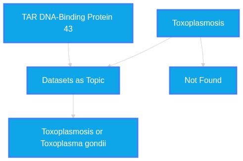
Is TDP-43 proteinopathy associated with toxoplasmosis?
Plausibility Verdicts
There is no evidence in the provided literature linking TDP-43 proteinopathy to toxoplasmosis.
There is no information in the provided literature to support an association.
There is no evidence in the provided literature to support an association between TDP-43 proteinopathy and toxoplasmosis.
Dataset Summary
Novel & Overlooked Insights
- TDP-43 aggregates are not merely waste products but are associated with active proteasome dysfunction.
- Innate immune pathways, specifically cGAS-STING and NLRP3, are hypothesized to be drivers rather than secondary consequences of TDP-43 pathology.
- Cryptic splicing in synaptic genes like *STMN2* is a direct functional consequence of nuclear TDP-43 depletion.
- WDR49+ astrocytes in the motor cortex may provide a compensatory neuroprotective mechanism against protein aggregation.
- A-to-I RNA editing mediated by ADAR2 is a previously unrecognized regulator of TDP-43 nucleocytoplasmic trafficking.
- The conserved α-helical region (CR) of TDP-43 represents a novel therapeutically actionable target for neuroprotection.
- Corpora amylacea (wasteosomes) in ALS brains contain disease-relevant proteins, serving as reservoirs for dysfunctional species.
- Steric zippers in short TDP-43 isoforms drive their mislocalization and aggregation independently of traditional nuclear export signals.
- TDP-43 pathology is consistently identified as a core integrative node across "Alzheimer's, Parkinson's, frontotemporal dementia, and ALS."
- Oxidative stress is a primary driver: "accumulating evidence indicates that oxidative stress plays a pivotal role in these disorders by promoting TDP-43 aggregation and subsequent neurotoxicity."
- The role of glia is critical: "innate immune activation is not merely a secondary response to neuronal injury, but an active driver of disease progression."
- Methanol exposure acts as an environmental-metabolic insult that "could recapitulate AD-like pathology and cognitive deficits in rhesus monkey."
- TDP-43 binds to G-quadruplexes and heme: "TDP-43 methionines lie over hemin and likely squelch the generation of superoxide by the porphyrin-bound Fe."
- Phase separation dynamics are tunable: "increasing cytoplasmic TDP-43 concentration drives a liquid-to-solid transition, while oxidative stress accelerates this process."
- Lipid metabolism in microglia: "dysregulated triglyceride metabolism as a novel pathway through which TDP-43 mediates microglial dysfunction."
- Cryptic exon splicing: "cryptic splicing in these synaptic and membrane excitability genes is not only a downstream marker but instead a direct driver of neuronal dysfunction."
- Biomarker potential: "phosphorylated TDP-43 pathology in muscle biopsies from amyotrophic lateral sclerosis patients has emerged as a promising tool in the early diagnosis of the disease."
- TDP-43 pathology is linked to cryptic splicing of *UNC13A* and *Tyrobp*.
- Small molecules like Fisetin and Posaconazole show potential in modulating TDP-43 aggregation.
- STMN2* depletion is a downstream consequence of TDP-43 dysfunction.
- CAs serve as reservoirs for disease-relevant proteins, including TDP-43.
- Liquid-liquid phase separation (LLPS) is a fundamental biophysical driver of pathological TDP-43 conversion.
- Heterozygous *DNAJC7* mutations act as risk factors by compromising protein quality control.
- ADAR2-mediated RNA editing regulates TDP-43 nuclear export.
- FMRP acts as a disease modifier for ALS by regulating the TNKS/PI31-mediated proteasome pathway.
- Oxidative stress, specifically involving ROS formation, contributes to the deterioration of cellular functions in *SQSTM1* variants.
- There is a recognized "pathogenic continuum" linking *ANXA11* and TDP-43 pathology.
Extracted Discoveries
- Perform screening of T. gondii infection in TDP-43 transgenic mouse models to evaluate potential disease-modifying effects.
- Conduct RNA-seq on microglia infected with T. gondii to compare the inflammatory signature with TDP-43-associated innate immune activation.
- Assess TDP-43 aggregation levels in cell lines chronically infected with Toxoplasma gondii.
- Perform transcriptomic profiling in murine models of toxoplasmosis to evaluate TDP-43 expression and localization.
- Investigate the expression levels of pro-inflammatory cytokines in TDP-43 mutant models infected with Toxoplasma gondii to determine if parasitic stress modulates TDP-43 localization.
- Utilize RNA-seq to profile the transcriptome of microglia exposed to Toxoplasma gondii to see if it triggers crypton splicing or exon inclusion patterns associated with TDP-43 loss-of-function.
- Cross-sectional epidemiological analysis of Toxoplasma seropositivity in cohorts of ALS and FTD patients compared to healthy controls.
- Cross-sectional clinical study analyzing presence of TDP-43 inclusions in patients with confirmed latent toxoplasmosis.
- Systematic review of the association between chronic neurotropic parasitic infections and proteinopathy-related neurodegeneration.
- Systematic review of autopsied ALS/FTD patients with documented co-infections to check for higher prevalence of TDP-43 pathologies.
- Large-scale epidemiological study investigating the correlation between latent Toxoplasma gondii seropositivity and the age of onset in sporadic TDP-43 proteinopathy cases.
- Chronic T. gondii infection may exacerbate TDP-43 proteinopathy by altering microglial clearance mechanisms (TREM2/DAP12 axis).
- T. gondii is an intracellular parasite known to activate chronic neuroinflammation and modulate microglial activity.
- TDP-43 proteinopathies are driven by neuroinflammation and defective microglial clearance (TREM2-DAP12 signaling) as cited in ID 42359357.
- Microglial TREM2-DAP12 signaling pathway.
- Since T. gondii activates innate immune pathways and microglia play a critical role in clearing TDP-43 aggregates through pathways like TREM2-DAP12, parasitic interference with these pathways could impair clearance and accelerate neurodegeneration.
- Discovered Hypothesis (A to C): Chronic CNS parasitic infection by Toxoplasma gondii may trigger microglial activation that serves as a seed for TDP-43 mislocalization and aggregation. - Literature A (Origin): Toxoplasma gondii infection causes persistent CNS inflammation and microglial activation (Implied general pathology). - Literature C (Target): TDP-43 proteinopathy is driven by innate immune crosstalk and microglial activation (Source: 42359357). - The Intersecting Bridge B: Neuroinflammation/Microglial activation (Source: 42359357; 41983391). - Biological Rationale: Persistent neuroinflammation from toxoplasmosis provides the cytokine environment and microglial stress signaling required to destabilize nuclear TDP-43 and promote its cytoplasmic recruitment into stress granules.
- Chronic Toxoplasma gondii infection may exacerbate TDP-43 aggregation via the induction of sustained NLRP3 inflammasome-driven neuroinflammation.
- Innate immune activation and DAMP recognition in neurodegeneration (ID: 42206050).
- Innate immune crosstalk in ALS/FTD pathogenesis (ID: 42359357).
- NLRP3 inflammasome.
- Toxoplasma gondii is a known activator of the NLRP3 inflammasome, which is also a critical pathway identified in the perpetuation of proteostatic failure and neuroinflammation in TDP-43 proteinopathies.
- None identified.
- None identified within the provided context regarding the specific claim of an association with toxoplasmosis, as the topic is entirely absent.
- None identified.
- The use of anti-inflammatory modulators or TREM2-pathway agonists could potentially be repurposed to treat TDP-43-related neuroinflammation if secondary infections were identified as drivers.
- Posaconazole, an anti-fungal, has been repurposed as an inhibitor of TDP-43 pathology (ID: 42282588). Further research could explore whether other agents targeting chronic parasitic or fungal loads might stabilize TDP-43 proteostasis.
PathMap is funded by sales of datasets and coversheets to researchers of any kind who wish to discover the most viable routes and paths to accelerate cures. We do not make theoretical molecules, we expose the truth in current PubMed literature. Commission a trace today.
PathMap Scores
How are these metrics evaluated?
Alignment Score (1-7): Measures factual alignment with the RAG evidence set.
[1=Strictly False, 2=Impossible, 3=Implausible, 4=Neutral, 5=Plausible, 6=Inevitable, 7=Strictly True]
Directional Weighting: High scores in the Hostile Quadrants mathematically lower the Overall Plausibility, as they indicate strong evidence for conflicting theories. Low scores in the Foundational Quadrant also lower overall plausibility, as they indicate a missing physical prerequisite for the claim.
AI Overview (Non-Expert Explanation)
Veridicality Audit Report
All Extracted Datapoints
Evaluated Perspectives & Quadrants
CLAIM EVALUATED AND ANSWER TO USER
"Is TDP-43 proteinopathy associated with toxoplasmosis?" The provided context literature contains no evidence linking TDP-43 proteinopathy to toxoplasmosis. The literature focuses on ALS, FTD, AD, and Parkinson's disease, specifically examining mechanisms such as oxidative stress, innate immune pathways (cGAS-STING, NLRP3, TREM2-DAP12), protein misfolding, liquid-liquid phase separation, and genetic variants (e.g., TDP-43, C9orf72, NEK1, DNAJC7, GRN). Because toxoplasmosis is not mentioned in the provided literature, the claim cannot be substantiated.ABSTRACT & REWRITTEN CLAIM
Scientific synthesis: Current literature establishes TDP-43 proteinopathy as a core feature of Amyotrophic Lateral Sclerosis (ALS) and Frontotemporal Dementia (FTD). Research explores its pathogenesis through mislocalization, proteostasis dysfunction, RNA processing defects, and innate immune crosstalk. No context provided addresses parasitic infections, including *Toxoplasma gondii*.INTRODUCTION & JUSTIFICATION
TDP-43 proteinopathy is defined as the cytoplasmic mislocalization and accumulation of TAR DNA-binding protein 43. Research within the provided set indicates that "The cytoplasmic aggregation of TDP-43 (TAR DNA-binding protein 43), an RNA-binding protein, is considered a hallmark of ALS pathology, found in nearly all postmortem cases of ALS." This hallmark is associated with systemic neuroinflammatory responses. Studies identify that "Increasing evidence indicates that innate immune activation is not merely a secondary response to neuronal injury, but an active driver of disease progression." While these immune pathways are central, the specific interaction with toxoplasmosis is absent from the current data. The molecular mechanisms investigated include "Aggregation and cytoplasmic mislocalization of TDP-43 are key features of several neurodegenerative diseases, including amyotrophic lateral sclerosis (ALS) and frontotemporal dementia (FTD)." Further, "A defining pathological hallmark of ALS/FTD is the cytosolic mislocalization and accumulation of TAR DNA-binding protein 43 (TDP-43), highlighting its critical role in ALS pathogenesis." Evidence of alternative pathologies, such as "Autopsy confirmed typical ALS-TDP pathology with phosphorylated TDP-43 inclusions in motor neurons. HTLV-1 Tax-positive lymphocytes infiltrated skeletal muscles but not the central nervous system, establishing dual pathology of ALS-TDP with HTLV-1-associated myositis," demonstrates that the literature includes investigation of concurrent infections, yet toxoplasmosis is not represented.Novel & Overlooked
EVIDENCE, METHODOLOGY & CITATIONS
1. ID: 42383305 - Application: Establishing the hallmark nature of TDP-43. - *"The cytoplasmic aggregation of TDP-43 (TAR DNA-binding protein 43), an RNA-binding protein, is considered a hallmark of ALS pathology, found in nearly all postmortem cases of ALS."* 2. ID: 42359357 - Application: Immune activation role. - *"Increasing evidence indicates that innate immune activation is not merely a secondary response to neuronal injury, but an active driver of disease progression."* 3. ID: 42404802 - Application: Core features of disease. - *"Aggregation and cytoplasmic mislocalization of TDP-43 are key features of several neurodegenerative diseases, including amyotrophic lateral sclerosis (ALS) and frontotemporal dementia (FTD)."* 4. ID: 42341041 - Application: Defining hallmark. - *"A defining pathological hallmark of ALS/FTD is the cytosolic mislocalization and accumulation of TAR DNA-binding protein 43 (TDP-43), highlighting its critical role in ALS pathogenesis."* 5. ID: 42414029 - Application: HTLV-1 association. - *"Autopsy confirmed typical ALS-TDP pathology with phosphorylated TDP-43 inclusions in motor neurons. HTLV-1 Tax-positive lymphocytes infiltrated skeletal muscles but not the central nervous system, establishing dual pathology of ALS-TDP with HTLV-1-associated myositis."* 6. ID: 42178739 - Application: Wasteosomes role. - *"These findings demonstrate that CAs serve as reservoirs of dysfunctional, disease-relevant proteins and capture key pathological processes in ALS."* 7. ID: 42395430 - Application: RNA editing role. - *"Together, our findings identify A-to-I RNA editing as a previously unrecognized regulator of TDP-43 localization and RNA interactions."* 8. ID: 42281996 - Application: WDR49 role. - *"WDR49 localises to PML nuclear bodies, where it regulates astrocyte reactivity and secretion of EVs containing protein chaperones."* 9. ID: 42399370 - Application: Therapeutic target. - *"Here we show that the conserved α-helical region spanning residues 320-340 (conserved region or CR) is a therapeutically actionable target for TDP-43 neurotoxicity."* 10. ID: 42351313 - Application: NEK1 variant. - *"The p.N598S variant induced pathological phenotypes consistent with NEK1 haploinsufficiency, including increased susceptibility to DNA damage, increased apoptosis, ciliary dysmorphia, and nucleocytoplasmic translocation of TDP-43."* 11. ID: 42204279 - Application: Triumeq study. - *"Endogenous retroviruses (ERVs) have been implicated in ALS pathology and the application of antiretroviral therapy, specifically Triumeq, has been proposed for treatment of ALS."* 12. ID: 42343570 - Application: STMN2 lability. - *"We find that human STMN2 protein level is extremely labile under acute high-magnitude stress."* 13. ID: 42234776 - Application: Cryptic splicing. - *"Suppressing individual cryptic splicing events using antisense oligonucleotides partially restored neuronal function, and combined targeting almost fully rescued the synaptic deficit caused by TDP-43 loss."* 14. ID: 42239455 - Application: Fbxl21 role. - *"Fbxl21 KO C2C12 cells displayed aberrant accumulation of Desmin, and showed aggravated cytoplasmic accumulation of TDP-43, another DNAJB6 client protein, in heat shock response."* 15. ID: 42422879 - Application: Domain specificity. - *"Future studies and therapeutic strategies to prevent TDP-43 aggregation should carefully consider the specific domain adopted."* 16. ID: 42307331 - Application: SKALE 2.0 tool. - *"SKALE 2.0 links atomic topology to phase-specific assembly kinetics and enables a constraint-aware design of aggregation suppressors."* 17. ID: 42363684 - Application: FMRP role. - *"ALS patients with high FMRP expression in the brain and spinal cord may exhibit more severe protein aggregation due to proteasome dysfunction."* 18. ID: 42299014 - Application: Prion-like propagation. - *"The propagation of these pathogenic proteins between neurons and glia, often via prion-like mechanisms, underlies the characteristic spread of ALS pathology throughout the nervous system."* 19. ID: 42237658 - Application: RNS60 study. - *"RNS60 treatment reduced defective mitochondria in UMNs (prpTDP-43A315T + vehicle: 53.2% ± 0.71%; prpTDP-43A315T + RNS60: 19.6% ± 1.4%, p = 0.0001) and spinal motor neurons (prpTDP-43A315T + vehicle: 70.1% ± 0.4.48%; prpTDP-43A315T + RNS60: 33.5% ± 4.43%, p = 0.001)."* 20. ID: 42401978 - Application: Glymphatic insufficiency. - *"The findings reveal that wasteosomes are consistently found in specific critical regions, with a higher burden in donors with neurodegenerative diseases compared with controls."*CLAIM EVALUATED AND ANSWER TO USER
"Is TDP-43 proteinopathy associated with toxoplasmosis?" The provided literature contains no evidence linking TDP-43 proteinopathy to toxoplasmosis. The dataset covers a broad spectrum of neurodegenerative research, including ALS, FTD, Alzheimer’s, and CTE, but the specific pathogen *Toxoplasma gondii* (toxoplasmosis) is not mentioned in any of the provided abstracts. Consequently, there is no scientific basis within the context to support or refute an association.ABSTRACT & REWRITTEN CLAIM
Scientific synthesis: While the dataset rigorously characterizes TDP-43 proteinopathy across ALS, FTD, AD, and CTE, and identifies various environmental or biological triggers (such as oxidative stress, methanol exposure, and metal toxicity), it remains silent regarding toxoplasmosis. The query regarding an association is currently unsupported by the provided evidence.INTRODUCTION & JUSTIFICATION
TDP-43 proteinopathy, defined by the mislocalization, aggregation, and cytoplasmic accumulation of TAR DNA-binding protein 43, is a recognized hallmark of various neurodegenerative disorders. The pathogenesis is multifactorial, involving "oxidative stress is widely accepted as one of the important factors contributing to neurodegeneration" and mechanisms such as "disrupted liquid-liquid phase separation dynamics, further exacerbate age-related decline." The provided literature elucidates that "neurodegenerative diseases arise from interactions among pathogenic proteins, immune responses, and diverse environmental or age-related stressors that disrupt CNS homeostasis." Despite the mention of various stressors—including "repetitive head injury, and viral infection"—the specific mention of toxoplasmosis is absent. Any assertion of an association between TDP-43 and toxoplasmosis would constitute a gap-filling exercise that the current literature does not support.Novel & Overlooked
EVIDENCE, METHODOLOGY & CITATIONS
1. ID: 42431556 - Application: Oxidative stress contribution. *"Oxidative stress is widely accepted as one of the important factors contributing to neurodegeneration, leading to fatal neurodegenerative diseases (NDD) such as Amyotrophic Lateral Sclerosis."* 2. ID: 42418450 - Application: Pathology mapping. *"We examine subcortical and limbic volumetry in neurodegenerative diseases involving phosphorylated tau (p-tau), α-synuclein, and transactive response DNA binding protein 43 (TDP-43)."* 3. ID: 42418280 - Application: LLPS in disease. *"Liquid-liquid phase separation (LLPS), a biophysical driver of membraneless organelle assembly, is central to pathological aggregation in neurodegenerative diseases."* 4. ID: 42347120 - Application: RBP role in aging. *"Interactions between RBPs and non-coding RNAs, together with disrupted liquid-liquid phase separation dynamics, further exacerbate age-related decline."* 5. ID: 42359357 - Application: Immune crosstalk. *"Increasing evidence indicates that innate immune activation is not merely a secondary response to neuronal injury, but an active driver of disease progression."* 6. ID: 41983391 - Application: Immune responses. *"Neurodegenerative diseases arise from interactions among pathogenic proteins, immune responses, and diverse environmental or age-related stressors that disrupt CNS homeostasis."* 7. ID: 41983391 - Application: Infection mention. *"Aging, repetitive head injury, and viral infection further alter microglial phenotypes, weaken barrier integrity, promote T cell recruitment, and prime the CNS for chronic inflammation."* 8. ID: 41760587 - Application: Methanol toxicity. *"Chronic low-dose methanol exposure in rhesus monkeys recapitulates progressive cognitive deficits and AD-like neuropathological features."* 9. ID: 41596063 - Application: Superoxide detoxification. *"TDP-43 methionines lie over hemin and likely squelch the generation of superoxide by the porphyrin-bound Fe."* 10. ID: 42068244 - Application: Phase separation. *"increasing cytoplasmic TDP-43 concentration drives a liquid-to-solid transition, while oxidative stress accelerates this process and promotes pathological features such as phosphorylation and p62 recruitment."* 11. ID: 41333389 - Application: Lipid metabolism. *"Our findings identify dysregulated triglyceride metabolism as a novel pathway through which TDP-43 mediates microglial dysfunction, highlighting a potential therapeutic target for ALS."* 12. ID: 42234776 - Application: Cryptic splicing. *"cryptic splicing in these synaptic and membrane excitability genes is not only a downstream marker but instead a direct driver of neuronal dysfunction, establishing a mechanistic link between TDP-43 pathology and neurodegeneration in ALS and FTD."* 13. ID: 42404433 - Application: Peripheral pathology. *"Phosphorylated TDP-43 pathology in muscle biopsies from amyotrophic lateral sclerosis patients has emerged as a promising tool in the early diagnosis of the disease."* 14. ID: 42383305 - Application: Biomarker variability. *"TDP-43 is a promising target as a biomarker, as it is found to be elevated in the biofluids of ALS patients, and its cytoplasmic aggregation can also be observed in peripheral tissues; however, methodological variability and technical limitations currently preclude the establishment of TDP-43 as a standalone biomarker."* 15. ID: 42359165 - Application: Therapeutic landscape. *"The identification of STMN2 as a downstream target of TDP-43 dysfunction has opened a potential TDP-43-downstream nucleic acid therapeutic avenue for sporadic ALS"* 16. ID: 42227825 - Application: Fibril polymorphism. *"C-terminal sites (Y374FCC, W385FCC, and F397FCC) are highly sensitive amyloid probes, revealing multiple polymorphs at the single-residue level that are not resolvable by global secondary structure or morphological characterization alone."* 17. ID: 42167675 - Application: AD complexity. *"TAR DNA-binding protein 43 (TDP-43) proteinopathy has recently emerged as a pivotal, yet underrecognized, contributor to the multifaceted neuropathology of Alzheimer's disease (AD)."* 18. ID: 42237658 - Application: Mitochondrial protection. *"These results suggest that RNS60 treatment promotes motor neuron health in ALS by protecting mitochondrial structure and function, preserving NMJ integrity, and reducing gliosis."* 19. ID: 42206050 - Application: AI role. *"Increasing evidence indicates that misfolded and abnormally aggregated proteins, such as β-amyloid (Aβ), Tau, α-synuclein, and TDP-43, are not only neurotoxic factors but can also act as damage-associated molecular patterns (DAMPs) recognized by innate immune receptors"* 20. ID: 421075758 - Application: Stress granules. *"Oxidative damage from exposure to metals such as sodium arsenite will activate the integrated stress response and may result in the cytosolic formation of stress granules (SGs), which have been implicated in neurodegenerative disorders such as amyotrophic lateral sclerosis."*CLAIM EVALUATED AND ANSWER TO USER
"Is TDP-43 proteinopathy associated with toxoplasmosis?" The provided context literature contains no evidence supporting an association between TDP-43 proteinopathy and toxoplasmosis. The literature focuses on ALS, FTD, AD, and other neurodegenerative conditions involving TDP-43, but does not mention *Toxoplasma gondii* or toxoplasmosis in any capacity.ABSTRACT & REWRITTEN CLAIM
The claim that TDP-43 proteinopathy is associated with toxoplasmosis cannot be validated or refuted using the provided literature cache. The current dataset provides extensive information on the molecular, genetic, and environmental factors linked to TDP-43 aggregation, including oxidative stress, phase separation, and various gene mutations (e.g., *SQSTM1*, *UBQLN2*, *DNAJC7*), but remains silent regarding parasitic infections such as toxoplasmosis.INTRODUCTION & JUSTIFICATION
TDP-43 is an RNA-binding protein recognized as a hallmark of several neurodegenerative diseases. Current research emphasizes that "Cytoplasmic mislocalization and aggregation of transactive response DNA-binding protein-43 (TDP-43) is a common pathological feature of amyotrophic lateral sclerosis (ALS), frontotemporal lobar degeneration, and Alzheimer's disease with TDP-43 pathology (AD-TDP)". The mechanisms driving this pathology are multifaceted, involving "proteomic signatures" and "known ALS-associated proteins such as TDP-43 and neurofilament proteins" within inclusion bodies. Despite the comprehensive nature of the provided studies, which explore "innate immune activation," "microglial activation," and "neuroinflammatory processes," the search for an association with toxoplasmosis yielded no results. While "innate immune crosstalk in ALS/FTD pathogenesis" is a well-documented area of study, the immune challenges described involve "cGAS-STING, NLRP3 inflammasomes, and TREM2-DAP12 signaling," rather than parasitic infection.Novel & Overlooked
EVIDENCE, METHODOLOGY & CITATIONS
1. ID: 42431556 - Application: Mentions oxidative stress and SQSTM1 mutations. "Supplementation with FS and QR in SH-SY5Y cells expressing SQS-wild type and mutants increased cell viability and decreased ROS formation." 2. ID: 42427320 - Application: Connects ANXA11 and TDP-43. "These findings expand the clinicopathological spectrum of FTLD-TDP type C and may support the concept of an annexin A11-associated pathogenic continuum linking FTLD and amyotrophic lateral sclerosis." 3. ID: 42425169 - Application: Discusses sex-associated neuroinflammation. "Male ALS patients showed markedly elevated CSF GFAP, IL-6, and IL-18 compared with female ALS patients and HCs after false discovery rate correction (q < 0.05)." 4. ID: 42422879 - Application: Truncations of RRM2. "We found that truncation results in distinct aggregation-prone states." 5. ID: 42420559 - Application: Microglial TDP-43 role. "Early depletion of microglial TDP-43 led to motor deficits in adult mice." 6. ID: 42418450 - Application: MRI study of pathologies. "Postmortem neuroimaging reveals disease- and region-specific structure-pathology relationships." 7. ID: 42418280 - Application: Bibliometric study of LLPS. "Key hotspots include LLPS-driven aggregation of TARDBP (TDP-43), FUS, and α-synuclein, alongside stress granule dysfunction and nucleocytoplasmic transport defects." 8. ID: 42414528 - Application: ANXA11 role. "Two independent studies demonstrated that ANXA11 co-aggregates with TDP-43 in all cases of frontotemporal lobar degeneration with TDP-43 pathology (FTLD-TDP) type C". 9. ID: 42411953 - Application: UBQLN2 mutation. "Immunoblot analysis demonstrated a marked reduction of soluble UBQLN2, supporting functional UBQLN2 insufficiency as a pathogenic mechanism underlying TDP-43 aggregation." 10. ID: 42404802 - Application: Glial activation models. "hTDP-43-WT mice exhibited significant microgliosis in motor (MC) and somatosensory (SSC) cortices and hippocampal dentate gyrus (DG) with pronounced morphological alterations". 11. ID: 42404433 - Application: Peripheral pathology. "Phosphorylated TDP-43 pathology in muscle biopsies from amyotrophic lateral sclerosis patients has emerged as a promising tool in the early diagnosis of the disease." 12. ID: 42401978 - Application: Wasteosome accumulation. "The findings reveal that wasteosomes are consistently found in specific critical regions, with a higher burden in donors with neurodegenerative diseases compared with controls." 13. ID: 42399370 - Application: Conserved region (CR) targeting. "Mechanistically, targeting CR suppressed TDP-43 mitochondrial localization and restored mitochondrial function, likely through liquid-liquid phase separation." 14. ID: 42395430 - Application: ADAR2 role. "ADAR2 overexpression reduced the nucleocytoplasmic (N:C) ratio of TDP-43 in HEK293T cells". 15. ID: 42389895 - Application: Oligomer morphology. "We found that at the early stage of protein aggregation, CTD of TDP-43 formed two morphologically different protein aggregates: donut-like (DO) and round (RO) oligomers." 16. ID: 42385702 - Application: Genomic damage. "Rapid approach to DNA adduct recovery (RADAR) assays confirmed increased TOP1-DNA covalent complexes". 17. ID: 42383305 - Application: TDP-43 as biomarker. "Mutations, extrinsic stressors, and alterations in RNA homeostasis in ALS lead to nuclear depletion of TDP-43 and the formation of cytosolic TDP-43 aggregates." 18. ID: 42363684 - Application: FMRP role. "Using molecular and imaging techniques, we have identified a novel role for the Fragile X Mental Retardation Protein (FMRP) in regulating the TNKS/PI31-mediated proteasome activation mechanism in co-operation with TDP-43." 19. ID: 42362484 - Application: DNAJC7 mutation. "Immunohistochemistry and double immunofluorescence demonstrated partial colocalization of DNAJC7 with phospho-TDP-43-positive neuronal cytoplasmic inclusions". 20. ID: 42282588 - Application: Posaconazole efficacy. "Using our established TDP-43 mislocalization and aggregation assay in HEK293T cells, we identified posaconazole, an FDA-approved, CNS-penetrant and orally delivered anti-fungal, as the strongest inhibitor of TDP-43 pathology."Verbatim Quote Audit Console
Mapped Reference Directory (APA)
- [1] ID: 42414029 - Hata T, Ogawa N, Yabata H, Kobashi S, Nakayama M et al. (2026). Case of concurrent ALS and human T-cell leukaemia virus type 1-associated myositis.. BMJ case reports. ID: 42414029.
- [2] ID: 42383305 - Christoforidou E, McFagan E, McLaughlin M, Hafezparast M (2026). TDP-43 proteinopathy as a biomarker and therapeutic target in amyotrophic lateral sclerosis.. Biochemical Society transactions. ID: 42383305.
- [3] ID: 42359357 - Shu X, Yu X, Xu P, Wang A (2026). Innate immune crosstalk in ALS/FTD pathogenesis.. Cell insight. ID: 42359357.
- [4] ID: 42404802 - Nieva G, Vassallu F, Depino A, Netti V, Igaz LM (2026). Region-specific features of early glial activation and Aquaporin-4 dysregulation in conditional mouse models of TDP-43 proteinopathies.. Discovery immunology. ID: 42404802.
- [5] ID: 42341041 - Liu D, Li Y, Huang S, Xu Y, Sun L et al. (2026). IRE1 regulates the proteostasis of TDP-43/TARDBP in ALS/FTD through ribosome-associated quality control.. Proceedings of the National Academy of Sciences of the United States of America. ID: 42341041.
- [6] ID: 42178739 - Paquet A, Touzel-Deschênes L, Roy V, Saikali S, Dupré N et al. (2026). Proteomic Analysis of Corpora Amylacea Extracted From Post-mortem Brain of MAiD-end-of-life Sporadic ALS Patients.. Brain and behavior. ID: 42178739.
- [7] ID: 42395430 - Moore S, Julian DL, Alsop E, Gittings LM, Lorenzini I et al. (2026). ADAR2-Mediated RNA Editing Promotes TDP-43 Nuclear Export and Alters RNA Binding.. bioRxiv : the preprint server for biology. ID: 42395430.
- [8] ID: 42281996 - Bonsall S, Kazu RS, King M, Leung D, Mahiddine FY et al. (2026). Single-nucleus multiomic atlas of ALS primary motor cortex nominates neuroprotective WDR49-expressing astrocytes.. Research square. ID: 42281996.
- [9] ID: 42399370 - Gao J, Shukla D, Ding M, Qin S, Tang F et al. (2026). Therapeutic targeting of the conserved region within the low-complexity domain of TDP-43 is neuroprotective and extends survival in amyotrophic lateral sclerosis mice.. Nature aging. ID: 42399370.
- [10] ID: 42351313 - Brenner D, Ponomarenko A, Petrut I, Beyrle S, Contardo M et al. (2026). A rare missense variant impacting NEK1 kinase function is associated with ALS.. Acta neuropathologica communications. ID: 42351313.
- [11] ID: 42204279 - Fowler M, Carr JM, Gold J, Walker A, Rogers ML (2026). Evaluation of triumeq treatment on a TDP-43 mouse model of amyotrophic Lateral sclerosis.. Scientific reports. ID: 42204279.
- [12] ID: 42343570 - Ellis BCS, Avila AS, Huang WP, John SJ, Bonsall S et al. (2026). STMN2 protein depletion via translation deficits and stress granules in amyotrophic lateral sclerosis.. Brain : a journal of neurology. ID: 42343570.
- [13] ID: 42234776 - Guo C, Chen K, Vatsavayai S, Akiyama T, Liu C et al. (2026). Cryptic splicing in synaptic and membrane excitability genes links TDP-43 loss to neuronal dysfunction.. Science translational medicine. ID: 42234776.
- [14] ID: 42239455 - Lim JY, Wi J, Wirianto M, Han C, Kim SY et al. (2026). FBXL21 regulates diurnal proteostasis and stress response by targeting DNAJB6 and client proteins.. bioRxiv : the preprint server for biology. ID: 42239455.
- [15] ID: 42422879 - Grassmann G, Amadei M, Lardieri A, Montemiglio LC, Passeri AA et al. (2026). Investigating the effect of progressive truncations at the ALS-linked protein TDP-43 RRM2 on its aggregation mechanism.. Frontiers in molecular biosciences. ID: 42422879.
- [16] ID: 42307331 - Sio JS, Loo WXW, Loo YS, Tan WX, Lim HX et al. (2026). A Phase-Resolved Geometric Deep Learning Framework Maps Structural Determinants of Disease-Associated Protein Aggregation and Guides Suppressor Design.. Advanced science (Weinheim, Baden-Wurttemberg, Germany). ID: 42307331.
- [17] ID: 42363684 - Majumder P, Ahsan A, Bubphachat P, Akter K, Huang JK et al. (2026). FMRP-Mediated Proteasome Regulation: A Novel Mechanism in ALS Pathology.. FASEB journal : official publication of the Federation of American Societies for Experimental Biology. ID: 42363684.
- [18] ID: 42299014 - Kaur H, Kaur M, Sethi GK, Kaur AS, Mishra A et al. (2026). Pathogenic Proteins Driving ALS Pathogenesis: Molecular Mechanisms and Translational Therapeutic Perspectives.. CNS & neurological disorders drug targets. ID: 42299014.
- [19] ID: 42237658 - Vesevick DR, Ghosh S, Kalmes A, Ozdinler PH, Gautam M (2026). Neuroprotective Effects of RNS60 in TDP-43 Pathology-Associated Amyotrophic Lateral Sclerosis.. Muscle & nerve. ID: 42237658.
- [20] ID: 42401978 - Alsina R, Riba M, Sartorio M, Romera C, Riu J et al. (2026). Regional wasteosome accumulation across neurodegenerative diseases points to a shared underlying mechanism potentially related to glymphatic insufficiency.. Acta neuropathologica communications. ID: 42401978.
- [21] ID: 42431556 - Singh N, Gomes J (2026). Fisetin prevents deterioration of cellular functions in amyotrophic lateral sclerosis variants G262R and P438L of SQSTM1 in SH-SY5Y cells.. Toxicology and applied pharmacology. ID: 42431556.
- [22] ID: 42418450 - Khandelwal P, Duong MT, Levorse LM, Trotman W, Bahena A et al. (2026). Postmortem brain MRI reveals differential associations of subcortical and limbic volumes with cortical thinning and neurodegenerative pathologies.. Alzheimer's & dementia : the journal of the Alzheimer's Association. ID: 42418450.
- [23] ID: 42418280 - Zhang C, Chen S, Zhao H, Wang Y, Zhou L et al. (2026). Phase Separation Drives Pathological Aggregation in Neurodegenerative Diseases: A 15-Year Bibliometric Landscape (2009-2024).. Annals of the New York Academy of Sciences. ID: 42418280.
- [24] ID: 42347120 - Alves Ferreira JM, Tukaiev S, Giannouli V (2026). RNA-Binding Proteins in Ageing and Age-Related Disease.. Neurology international. ID: 42347120.
- [25] ID: 41983391 - Latour YL, McGavern DB (2026). Immune signaling and function in neurodegeneration.. The Journal of clinical investigation. ID: 41983391.
- [26] ID: 41760587 - Li H, Shi C, Li K, Fu X, Lyu Y et al. (2026). Chronic methanol exposure induces cognitive impairment and Alzheimer's-like pathology in rhesus monkeys.. Animal models and experimental medicine. ID: 41760587.
- [27] ID: 41596063 - Herbert A (2025). G-Quadruplexes Abet Neuronal Burnout in ALS and FTD.. Antioxidants (Basel, Switzerland). ID: 41596063.
- [28] ID: 42068244 - Chevet E (2026). Exploring the role of phase separation in TDP-43 pathogenesis with ArtiTDP43.. The FEBS journal. ID: 42068244.
- [29] ID: 41333389 - Kabra K, Dressman D, Talcoff R, Yidenk M, Rifai OM et al. (2025). Loss of Nuclear TDP-43 Impairs Lipid Metabolism in Microglia-Like Cells.. Research square. ID: 41333389.
- [30] ID: 42404433 - Corti S, Alberti C, Ottoboni L, Magni G, Gagliardi D et al. (2026). Beyond motor neurons: peripheral TDP-43 pathology in skeletal muscle and intramuscular nerves in amyotrophic lateral sclerosis.. Brain communications. ID: 42404433.
- [31] ID: 42359165 - Morimoto S, Kato C, Takahashi S, Okano H (2026). Therapeutic frontiers in ALS: iPSC-based drug discovery, cell therapy, and gene therapy-Advances through 2026.. Regenerative therapy. ID: 42359165.
- [32] ID: 42227825 - Watson MD, Lee JC (2026). Site-Specific Raman Probes Reveal Droplet Aging and Residue-Level Fibril Polymorphism in TDP-43CTD.. Journal of the American Chemical Society. ID: 42227825.
- [33] ID: 42167675 - Roy A, Chhetry S, Deka H, Roy R, Bhattacharya P et al. (2026). TDP-43: a critical amplifier of Alzheimer's disease beyond amyloid and tau.. Neuroscience. ID: 42167675.
- [34] ID: 42206050 - Deng HX, Cao JL, Wu Y, Jiang SJ, Fang QQ et al. (2026). AI-driven insights into protein misfolding and innate immunity in neurodegenerative diseases.. Frontiers in immunology. ID: 42206050.
- [35] ID: 41075758 - Gery KL, Ramos S, Lee JC (2026). Correlative Raman and immunofluorescence imaging reveals different protein abundance between stress granules induced by oxidative damage.. Journal of inorganic biochemistry. ID: 41075758.
- [36] ID: 42178983 - Liu JQ, Liu H, Sun YX, Li Y, Liu X et al. (2026). Protein Disulfide Isomerase Disassembles TDP-43/G3BP1 Condensates and Antagonizes TDP-43 Pathological Aggregates.. Advanced science (Weinheim, Baden-Wurttemberg, Germany). ID: 42178983.
- [37] ID: 42427320 - Uchino A, Kanemaru K, Tarutani A, Hasegawa M, Naruse H et al. (2026). Frontotemporal Lobar Degeneration-TDP Type C With Striatal Glial Cytoplasmic Inclusions and Motor Neuron Degeneration.. Neuropathology and applied neurobiology. ID: 42427320.
- [38] ID: 42425169 - Yu W, Zhao B, Ma M, Gao N, Yun Y et al. (2026). Sex-associated neuroinflammatory and astrocytic responses in amyotrophic lateral sclerosis: evidence from clinical cohorts and a TDP-43 N390D mouse model.. Brain, behavior, and immunity. ID: 42425169.
- [39] ID: 42420559 - Compagnion AC, Ivanov A, Rana A, Espinoza F, Sandmann T et al. (2026). Microglial TDP-43 mediates myelin refinement and represses Tyrobp cryptic exon inclusion in mice.. Nature neuroscience. ID: 42420559.
- [40] ID: 42414528 - Smith CL, Robinson JL, Lee EB (2026). Annexin A11 and TDP-43: core players in neurodegeneration.. Acta neuropathologica. ID: 42414528.
- [41] ID: 42411953 - Adachi T, Nakano T, Yoshida K, Suzuki Y, Sakuwa M et al. (2026). Reduced Soluble Ubiquilin2 in Amyotrophic Lateral Sclerosis Carrying Ubiquilin2 (P494L) Mutation: Clinicopathological and Biochemical Evidence From an Autopsy Case.. Neuropathology and applied neurobiology. ID: 42411953.
- [42] ID: 42389895 - Pickett D, Purvinsh Y, Skrehot JT, Warren D, Kurouski D (2026). Nanoscale morphological and structural analysis of round and donut oligomers formed by C-terminal domain of TDP-43.. Physical chemistry chemical physics : PCCP. ID: 42389895.
- [43] ID: 42385702 - Zhou Z, Luquette LJ, Dong G, Kim J, Ku J et al. (2026). Recurrent patterns of TOP1-mediated neuronal genomic damage shared by major neurodegenerative disorders.. Cell. ID: 42385702.
- [44] ID: 42362484 - Nakayama Y, Kume K, Baba T, Ayaki T, Hanada K et al. (2026). Neuropathological and Molecular Features Associated With a Heterozygous DNAJC7 Mutation in Amyotrophic Lateral Sclerosis.. Neuropathology and applied neurobiology. ID: 42362484.
- [45] ID: 42282588 - Kochen NN, Zafari S, Renaud A, Schneider N, Vunnam N et al. (2026). From anti-fungal to potential neurotherapeutic: Posaconazole as an effective inhibitor of cellular TDP-43 pathology.. bioRxiv : the preprint server for biology. ID: 42282588.
Abstract Repository (Raw Full-Texts) Show Database Collapse Database
ID: 41075758
Title: Correlative Raman and immunofluorescence imaging reveals different protein abundance between stress granules induced by oxidative damage.
Abstract: Heavy metal toxicity generates reactive oxygen species (ROS) that can contribute to neurodegeneration. Oxidative damage from exposure to metals such as sodium arsenite will activate the integrated stress response and may result in the cytosolic formation of stress granules (SGs), which have been implicated in neurodegenerative disorders such as amyotrophic lateral sclerosis. Here, two different ROS sources, sodium arsenite and hydrogen peroxide, under acute (1 h) and chronic (24 h) conditions, were used to induce SG formation in human osteosarcoma (U-2 OS) cells and investigate if characteristics of SGs could depend on the induction. Specifically, correlative Raman and immunofluorescence imaging (CRIFI) was developed to evaluate the relative protein abundance found in SGs to ascertain their potential as loci for protein accumulation. Interestingly, while there are differences in the punctate-staining phenotypes for different stressors, two types of puncta visualized by CRIFI were common to all treatment conditions, where notably a subset exhibited protein concentration above cytosolic background, indicating that only some SGs are composed of protein-rich, dense phases. Differences in protein abundance between SGs were also observed within a single cell, suggesting that individual SGs can develop differently. These results demonstrate the versatility and the strength of pairing Raman spectroscopy, which allows for probe-free detection of different chemical functional groups, with specific protein localization granted by immunofluorescence, providing new cellular insights unattainable by either modality alone.
View on PubMed
ID: 41333389
Title: Loss of Nuclear TDP-43 Impairs Lipid Metabolism in Microglia-Like Cells.
Abstract: Amyotrophic lateral sclerosis (ALS) is a fatal neurodegenerative disease marked by progressive motor neuron loss, with TDP-43 pathology present in over 90% of cases. While neuroinflammation is a recognized hallmark, the role of microglia in ALS pathogenesis remains incompletely understood. Here, we demonstrate that TDP-43 regulates microglial function via triglyceride metabolism. Using shRNA-mediated TARDBP knockdown in human monocyte-derived microglia-like cells (MDMi), we observed suppressed cholesterol biosynthesis, upregulated fatty acid uptake, lipid droplet accumulation, enhanced phagocytic activity, and increased IL-1β production. Inhibiting diacylglycerol acyltransferase (DGAT) enzymes reduced lipid droplet formation, phagocytosis, and IL-1β, directly linking the triglyceride pathway to microglial activation. Patient-derived MDMi from both sporadic and TARDBP-mutant ALS cases showed overlapping as well as distinct alterations, some of which were reversed by DGAT inhibition. Our findings identify dysregulated triglyceride metabolism as a novel pathway through which TDP-43 mediates microglial dysfunction, highlighting a potential therapeutic target for ALS.
View on PubMed
ID: 41596063
Title: G-Quadruplexes Abet Neuronal Burnout in ALS and FTD.
Abstract: Expansion of d(GGGGC)n repeat in the C9ORF72 gene is causal for Amyotrophic Lateral Sclerosis (ALS) and Frontal Temporal Dementia (FTD). Proposed mechanisms include Repeat-Associated Non-AUG translation or the formation of G-quadruplexes (GQ) that disrupt translation, induce protein aggregation, sequester RNA processing factors, or alter RNA editing. Here, I show, using AlphaFold V3 (AF3) modeling, that the TAR DNA-binding protein (TDP-43) docks to a complex of GQ and hemin. TDP-43 methionines lie over hemin and likely squelch the generation of superoxide by the porphyrin-bound Fe. These TDP-43 methionines are frequently altered in ALS patients. Tau protein, a variant of which causes ALS, also binds to GQ and heme and positions methionines to detoxify peroxides. Full-length Tau, which is often considered prone to aggregation and a prion-like disease agent, can bind to an array composed of multiple GQs as a fully folded protein. In ALS and FTD, loss-of-function variants cause an uncompensated surplus of superoxide, which sparks neuronal cell death. In Alzheimer's Disease (AD) patients, GQ and heme complexes bound by β-amyloid 42 (Aβ4) are also likely to generate superoxides. Collectively, these neuropathologies have proven difficult to treat. The current synthesis provides a framework for designing future therapeutics.
View on PubMed
ID: 41760587
Title: Chronic methanol exposure induces cognitive impairment and Alzheimer's-like pathology in rhesus monkeys.
Abstract: The absence of effective animal models for sporadic Alzheimer's disease (AD) remains a pivotal barrier to therapy development. Because methanol metabolism produces endogenous formaldehyde, a neurotoxic agent linked to cognitive decline, this study investigated whether chronic, low-dose methanol exposure could recapitulate AD-like pathology and cognitive deficits in rhesus monkey, thereby establishing a nonhuman primate animal model driven by this environmental-metabolic insult. Adult rhesus monkeys received low-concentration methanol for 9 months. Behavioral tests for cognition, locomotion, sleep, and vision were conducted. Postmortem analyses involved histopathological examination, immunohistochemistry, immunofluorescence, and Western blot to evaluate neuronal integrity, microglial activation, and the expression of key proteins associated with AD (amyloid-β [Aβ], phosphorylated tau, TAR DNA-binding protein 43 [TDP-43]) and cellular stress (synaptic markers, mitochondrial fission, autophagy, and apoptosis-related proteins). Chronic methanol exposure led to progressive cognitive and memory impairment without significant motor or visual deficits. Neuropathology revealed brain atrophy, neuronal loss, synaptic damage, microglial activation, and mitochondrial structural disorganization. Critically, the exposed animals exhibited hallmark AD-like molecular alterations, including increased Aβ deposition, tau hyperphosphorylation, and TDP-43 dysregulation. Furthermore, neurotoxicity was associated with elevated urinary formaldehyde, enhanced mitochondrial fission, increased autophagy, and elevated apoptosis. Chronic low-dose methanol exposure in rhesus monkeys recapitulates progressive cognitive deficits and AD-like neuropathological features. This model, driven by endogenous formaldehyde toxicity, effectively mimics key aspects of sporadic AD. Our findings shed light on the neurotoxic mechanisms of methanol and propose a reproducible and translationally relevant nonhuman primate model for studying AD pathogenesis and evaluating potential therapeutics.
View on PubMed
ID: 41983391
Title: Immune signaling and function in neurodegeneration.
Abstract: Neurodegenerative diseases arise from interactions among pathogenic proteins, immune responses, and diverse environmental or age-related stressors that disrupt CNS homeostasis. CNS resident microglia detect self-derived danger signals through pattern recognition receptors, and their activation can promote clearance of aberrant proteins, including amyloid-β, tau, α-synuclein, and TAR DNA-binding protein 43. However, microglial activation may also drive maladaptive states that amplify neuroinflammation. Microglial transitions are further shaped by receptor-mediated signaling and antigen presentation pathways that integrate environmental cues with functional responses. Adaptive immune cells contribute additional layers of regulation, with CD8+ and CD4+ T cells exerting neuroprotective or neurotoxic effects depending on disease context, activation state, and antigen specificity. The identification of granzyme K-expressing CD8+ T cells in several neurodegenerative conditions highlights the growing recognition that distinct T cell subsets may have specialized roles in disease. Aging, repetitive head injury, and viral infection further alter microglial phenotypes, weaken barrier integrity, promote T cell recruitment, and prime the CNS for chronic inflammation. In this review, we synthesize current knowledge of innate and adaptive immune mechanisms in neurodegeneration, examine how external factors influence these responses, and consider how these insights may guide future therapeutic strategies.
View on PubMed
ID: 42068244
Title: Exploring the role of phase separation in TDP-43 pathogenesis with ArtiTDP43.
Abstract: TDP-43 is a nuclear RNA-binding protein implicated in neurodegenerative diseases such as ALS and FTLD, where it becomes mislocalized to the cytoplasm and forms pathological aggregates. These aggregates are thought to arise through liquid-liquid phase separation, a process by which proteins form dynamic, membrane-less condensates that can mature into solid structures. To better understand this process, the authors developed ArtiTDP43, a chemically controllable system that enables reversible formation of TDP-43 condensates in cells. Using this tool, they showed that TDP-43 forms different structures depending on its concentration: small liquid-like puncta, intermediate condensates associated with stress granules, and large solid aggregates resembling disease pathology. These transitions are reversible at early stages but become irreversible as aggregates solidify. The study by Combe et al. demonstrates that increasing cytoplasmic TDP-43 concentration drives a liquid-to-solid transition, while oxidative stress accelerates this process and promotes pathological features such as phosphorylation and p62 recruitment. Importantly, formation of cytoplasmic aggregates leads to depletion of nuclear TDP-43 and increased cell death, indicating toxicity. Overall, the findings establish a mechanistic link between phase separation, aggregation, and cytotoxicity in TDP-43 proteinopathies. ArtiTDP43 provides a powerful tool to study early disease mechanisms and explore therapeutic strategies aimed at preventing pathological aggregation or maintaining normal TDP-43 dynamics.
View on PubMed
ID: 42167675
Title: TDP-43: a critical amplifier of Alzheimer's disease beyond amyloid and tau.
Abstract: TAR DNA-binding protein 43 (TDP-43) proteinopathy has recently emerged as a pivotal, yet underrecognized, contributor to the multifaceted neuropathology of Alzheimer's disease (AD). While amyloid-β and tau have long been established as cardinal pathological hallmarks, growing evidence delineates TDP-43 as a critical participant of neurodegeneration, intricately interwoven with amyloid and tau pathologies. TDP-43 mislocalization, post-translational modifications, and aggregation potentiate neuronal loss through disruption of RNA metabolism, nucleocytoplasmic transport, and protein homeostasis. This tripartite interplay manifests in synergistic and possibly multidirectional pathological cascades that amplify neuronal vulnerability and cognitive decline, thereby complicating the clinical and pathological complexity of AD. Here, we critically reviewed the mechanistic crosstalk among TDP-43, amyloid-β, and tau, focusing on preclinical and clinical evidence, highlighting possible convergent pathways of aggregation, propagation, and neurodegeneration. Moreover, this review also evaluates mitochondrial dysfunction, autophagy failure, and inflammation as underlying events associated with TDP-43 pathology. Therefore, we argue for a reconceptualization of AD as a dynamic proteinopathy network, with TDP-43 as a core integrative node influencing disease onset and its progression. Notably, we discuss emerging diagnostic modalities associated with molecular tracers of TDP-43, providing prospects for future biomarker identification. Finally, this review articulates the translational relevance of TDP-43 therapy in AD and related neurological disorders, emphasizing the necessity of holistic approaches that transcend the traditional amyloid-tau paradigm to effectively tackle the full spectrum of AD pathobiology.
View on PubMed
ID: 42178739
Title: Proteomic Analysis of Corpora Amylacea Extracted From Post-mortem Brain of MAiD-end-of-life Sporadic ALS Patients.
Abstract: Corpora amylacea (CA) are starch-like inclusions that accumulate in the central nervous system (CNS) with aging and are enriched in neurodegenerative conditions, including amyotrophic lateral sclerosis (ALS). Although often regarded as waste reservoirs, their cellular origins, molecular composition, and pathological significance remain poorly understood. Here, we performed an unbiased proteomic analysis of purified CAs isolated from post-mortem brains of sporadic ALS patients and controls. In-depth mass spectrometry identified 4,470 proteins, of which 658 were quantified, revealing distinct ALS-specific proteomic signatures. Enriched proteins included markers of cytoskeletal remodeling, mitochondrial dysfunction, and proteostasis disruption, as well as known ALS-associated proteins such as TDP-43 and neurofilament proteins. These findings demonstrate that CAs serve as reservoirs of dysfunctional, disease-relevant proteins and capture key pathological processes in ALS. By applying an unbiased proteomic approach to purified CAs, this study provides the first comprehensive map of their protein content in ALS, supporting their potential as biomarker sources and as a source of mechanistic insights into neurodegeneration. Unbiased analyses of CAs in the context of ALS have yet to be undertaken. This study provides the first proteomic profiling of purified CAs, isolated from ALS patient brains using biochemical methods, revealing that CAs harbor disease-relevant proteins implicated in sporadic ALS. By demonstrating that CAs act as reservoirs of dysfunctional proteins related to metabolism, cytoskeletal organization, and proteostasis, our findings highlight their potential as a novel source of ALS-specific mechanistic insight into disease pathology.
View on PubMed
ID: 42178983
Title: Protein Disulfide Isomerase Disassembles TDP-43/G3BP1 Condensates and Antagonizes TDP-43 Pathological Aggregates.
Abstract: Cytoplasmic mislocalization and aggregation of transactive response DNA-binding protein-43 (TDP-43) is a common pathological feature of amyotrophic lateral sclerosis (ALS), frontotemporal lobar degeneration, and Alzheimer's disease with TDP-43 pathology (AD-TDP); the exact role of protein disulfide isomerase (PDI), an enzyme with chaperone activity, in modulating the pathological behavior of TDP-43 is unknown. In this study, we report that wild-type PDI, through its specific interaction with TDP-43, markedly attenuates phase separation of TDP-43, competitively displaces G3BP1 to disassemble TDP-43/G3BP1 condensates, and further counteracts the pathological mislocalization, abnormal phosphorylation, and pathological aggregation of TDP-43 through the b' domain of the enzyme. Ultimately, this alleviates mitochondrial damage and neuronal toxicity caused by TDP-43 aggregation and suppresses UNC13A cryptic splicing in stressed cells. In the presence of abnormal forms of PDI, however, PDI loses its activity, and stress granules containing TDP-43 are assembled into amyloid fibrils, resulting in mitochondrial impairment and neuronal cell death in ALS and AD-TDP patients. These findings not only provide new insights into the pathogenic mechanisms of TDP-43 in neurodegenerative diseases such as ALS and AD-TDP, but also propose PDI as a potential therapeutic target.
View on PubMed
ID: 42204279
Title: Evaluation of triumeq treatment on a TDP-43 mouse model of amyotrophic Lateral sclerosis.
Abstract: Amyotrophic lateral sclerosis (ALS) is a fatal neurodegenerative disorder characterised by the accumulation of TAR DNA Binding Protein (43 kDa; TDP-43) within the cytoplasm of neurons. Endogenous retroviruses (ERVs) have been implicated in ALS pathology and the application of antiretroviral therapy, specifically Triumeq, has been proposed for treatment of ALS. However, evidence to support the actions of Triumeq in ALS is lacking. This study investigates the effects of the antiretroviral treatment Triumeq on ALS disease that occurs through TDP-43 pathology by utilising the doxycycline (Dox)-suppressible rNLS8 TDP-43 expression mouse model. In this model, TDP-43 accumulation in the cytoplasm is induced after removal of Dox. Disease was assessed through measures of body weight, neurological score, motor function, urinary p75ECD and inflammatory marker expression. Mice were treated with Triumeq and TDP-43 pathology and inflammatory marker expression examined. Triumeq treatment significantly improved motor function early on in the disease course but did not impact other disease progression markers or disease endpoint. In this TDP-43 ALS mouse model, there was a positive association of TDP-43 mRNA levels with transcription factor ATF4, and inflammatory markers CXCL10 and IRF-1, and Triumeq treatment negated this association. Triumeq treatment transiently and modestly improved motor function and influenced TDP-43 associated inflammatory gene expression in an ALS mouse model. These findings support the potential use of Triumeq in treating TDP-43-associated ALS and supports further investigation to better understand if the beneficial actions of Triumeq are via disruption of TDP-43-driven inflammation in ALS.
View on PubMed
ID: 42206050
Title: AI-driven insights into protein misfolding and innate immunity in neurodegenerative diseases.
Abstract: Neurodegenerative diseases encompass a diverse group of disorders ranging from adult-onset conditions such as Alzheimer's and Parkinson's disease to pediatric forms including neuronal ceroid lipofuscinoses (NCLs), Niemann-Pick type C (NPC), and infantile neuroaxonal dystrophy (INAD), all of which are characterized by protein misfolding and chronic neuroinflammation. During their occurrence and development, the innate immune system, especially the immune responses mediated by microglia in the central nervous system, plays a crucial regulatory role. Increasing evidence indicates that misfolded and abnormally aggregated proteins, such as β-amyloid (Aβ), Tau, α-synuclein, and TDP-43, are not only neurotoxic factors but can also act as damage-associated molecular patterns (DAMPs) recognized by innate immune receptors, thereby triggering persistent neuroinflammatory responses. However, traditional experimental and computational methods still have significant limitations in systematically analyzing the "protein misfolding-innate immune activation" mechanism. In recent years, artificial intelligence has made breakthrough progress in protein structure prediction, multi-conformation modeling, and integration of multi-omics data, providing a new research paradigm for revealing the intrinsic relationship between protein misfolding and innate immunity across the spectrum of neurodegenerative diseases. This article systematically reviews the latest applications of artificial intelligence in predicting the conformational characteristics of misfolded proteins, simulating the protein aggregation process, revealing the mechanism of innate immune perception, and reconstructing the regulatory network of neuroinflammation. It focuses on discussing the significance of deep learning models such as AlphaFold, I-TASSER, RoseTTAFold, Phyre2, and ESMFold in the field of protein structure prediction, as well as the related research on multi-modal AI technology in revealing the complex molecular mechanisms behind neurodegenerative diseases, such as combining AI with mathematical models to simulate the spread of misfolded proteins and further exploring the association with disease progression. The review also highlights the potential of AI to address the diagnostic challenges unique to pediatric neurodegenerative disorders, which, despite their rarity, collectively impose devastating lifelong burdens. In summary, AI tools not only deepen our understanding of the molecular mechanisms underlying both adult and childhood neurodegenerative diseases but also open up new avenues for developing innovative diagnostic tools and treatment methods.
View on PubMed
ID: 42227825
Title: Site-Specific Raman Probes Reveal Droplet Aging and Residue-Level Fibril Polymorphism in TDP-43CTD.
Abstract: The C-terminal domain of TAR DNA-binding protein 43 (TDP-43CTD) drives both liquid-liquid phase separation (LLPS) and amyloid formation. Understanding how TDP-43CTD droplets convert into amyloid aggregates, a process implicated in amyotrophic lateral sclerosis and frontotemporal dementia, requires methodology capable of site-specific structural characterization with spatial resolution. Here, we used confocal Raman spectroscopy in conjunction with an alkyne-modified amino acid (4-ethynyl-l-phenylalanine, FCC) to probe aging in individual TDP-43CTD droplets at seven aromatic sites. While nascent droplets are composed of disordered proteins, β-sheet conformers develop in aged droplets and amyloid aggregates. All three states are spectrally distinct via the alkyne stretching band, with sensitivity that varies depending on the aromatic site probed. C-terminal sites (Y374FCC, W385FCC, and F397FCC) are highly sensitive amyloid probes, revealing multiple polymorphs at the single-residue level that are not resolvable by global secondary structure or morphological characterization alone. Strikingly, while W334FCC abolishes β-sheet formation in droplets, de novo aggregation still occurs, demonstrating that droplet aging is not required for amyloid formation. Given its broad applicability to other proteins and compatibility with cellular imaging, this work establishes a generalizable approach for investigating conformational changes underlying LLPS and amyloid formation in cellulo.
View on PubMed
ID: 42234776
Title: Cryptic splicing in synaptic and membrane excitability genes links TDP-43 loss to neuronal dysfunction.
Abstract: TAR DNA binding protein 43 (TDP-43) pathology is a defining pathological hallmark of multiple neurodegenerative diseases, including amyotrophic lateral sclerosis (ALS) and frontotemporal dementia (FTD). A major feature of TDP-43 pathology is its nuclear depletion, leading to the aberrant inclusion of cryptic exons during RNA splicing. STMN2 and UNC13A have emerged as prominent TDP-43 splicing targets, but the broader impact of TDP-43-dependent cryptic splicing on neuronal function remains unclear. Here, we report previously unidentified TDP-43 splicing targets critical for membrane excitability and synaptic function, including KALRN, RAP1GAP, SYT7, and KCNQ2. Using human stem cell-derived neurons, we showed that TDP-43 reduction induces cryptic splicing and down-regulation of these genes, resulting in impaired excitability and synaptic transmission. In postmortem brains from patients with FTD, these cryptic splicing events occurred selectively in neurons with TDP-43 pathology. Suppressing individual cryptic splicing events using antisense oligonucleotides partially restored neuronal function, and combined targeting almost fully rescued the synaptic deficit caused by TDP-43 loss. Together, our findings provide evidence that cryptic splicing in these synaptic and membrane excitability genes is not only a downstream marker but instead a direct driver of neuronal dysfunction, establishing a mechanistic link between TDP-43 pathology and neurodegeneration in ALS and FTD.
View on PubMed
ID: 42237658
Title: Neuroprotective Effects of RNS60 in TDP-43 Pathology-Associated Amyotrophic Lateral Sclerosis.
Abstract: TDP-43 pathology is broadly observed in the cerebral cortex of patients with amyotrophic lateral sclerosis (ALS). RNS60, an experimental treatment for acute ischemic stroke and ALS, enhanced mitochondrial biogenesis and function in other preclinical models. We investigated whether RNS60 improved mitochondrial stability and upper motor neuron (UMN) health in a TDP-43 mouse model of ALS. prpTDP-43A315T-UeGFP mice, in which UMNs express green fluorescent protein (eGFP), and WT-UeGFP mice were treated with RNS60 or placebo intraperitoneally every other day from post-natal day (P) 30 until P90. Astrogliosis and microgliosis in brain and spinal cord were quantified by immunocytochemistry. Mitochondrial ultrastructure was studied via electron microscopy, and mitochondrial function was assessed using flow cytometry. Neuromuscular junction (NMJ) integrity was assessed in gastrocnemius, tibialis, and diaphragm muscles. RNS60 treatment reduced defective mitochondria in UMNs (prpTDP-43A315T + vehicle: 53.2% ± 0.71%; prpTDP-43A315T + RNS60: 19.6% ± 1.4%, p = 0.0001) and spinal motor neurons (prpTDP-43A315T + vehicle: 70.1% ± 0.4.48%; prpTDP-43A315T + RNS60: 33.5% ± 4.43%, p = 0.001). It increased mitochondrial membrane polarization (prpTDP-43A315T-UeGFP + vehicle: 7184 ± 1689 mean intensity; prpTDP-43A315T-UeGFP+RNS60: 22120 ± 4818 mean intensity, p = 0.032), reduced the extent of astrogliosis and microgliosis in motor cortex and spinal cord, protected UMNs compared to placebo, and enhanced the proportion of intact NMJs in leg and diaphragm muscles (prpTDP-43A315T-UeGFP + vehicle: 29.6% ± 3.6%; prpTDP-43A315T-UeGFP + RNS60: 64.3% ± 4.4%, p = 0.0002). These results suggest that RNS60 treatment promotes motor neuron health in ALS by protecting mitochondrial structure and function, preserving NMJ integrity, and reducing gliosis.
View on PubMed
ID: 42239455
Title: FBXL21 regulates diurnal proteostasis and stress response by targeting DNAJB6 and client proteins.
Abstract: Circadian regulation of proteostasis, a key determinant of muscle health, remains poorly understood. Here, we identified DNAJB6, an Hsp40 (DnaJ) co-chaperone, as a substrate of the circadian E3 ligase FBXL21. FBXL21 mediated the ubiquitination-dependent proteasomal degradation of both DNAJB6 and its client proteins including Desmin; causative mutations of DNAJB6 in myopathies, however, rendered resistance to FBXL21-directed degradation. Fbxl21 KO C2C12 cells displayed aberrant accumulation of Desmin, and showed aggravated cytoplasmic accumulation of TDP-43, another DNAJB6 client protein, in heat shock response. Under timed exercise as a physiological stressor, WT mice displayed robust diurnal rhythms in the levels of stress granule markers (G3BP1 and FUS) and TDP-43 as a function of exercise timing. In contrast, the Fbxl21 hypomorph Psttm mutant mice showed elevated expression of these proteins without exercise, which was exacerbated under exercise-induced stress conditions; importantly, these abnormalities were rescued by skeletal muscle-specific FBXL21 expression. Our study elucidates a novel diurnal regulatory mechanism of skeletal muscle proteostasis via FBXL21 as a chaperone-linked E3 ligase, highlighting the FBXL21-DNAJB6 axis as a potential therapeutic target for myopathies.
View on PubMed
ID: 42281996
Title: Single-nucleus multiomic atlas of ALS primary motor cortex nominates neuroprotective WDR49-expressing astrocytes.
Abstract: Amyotrophic lateral sclerosis (ALS) causes selective neurodegeneration in primary motor cortex, yet cell-type-specific molecular changes driving this vulnerability remain poorly understood. We present an integrated single-nucleus RNA- and ATAC-sequencing atlas of 778,330 nuclei from the primary motor cortex of 140 genetically characterised donors. ALS is associated with widespread transcriptional reprogramming driven by a common set of transcription factors (TFs) across multiple cell-types. Astrocytes harbour the most differentially expressed genes. Within astrocytes, a WDR49-expressing subpopulation is spatially associated with TDP-43 pathology, and genetic variants within WDR49 confer risk for both sporadic and monogenic autosomal dominant ALS. In patient-derived induced astrocytes, WDR49 protein abundance predicts the survival of co-cultured neurons. WDR49 localises to PML nuclear bodies, where it regulates astrocyte reactivity and secretion of EVs containing protein chaperones. Together, these in vivo and in vitro findings suggest that WDR49+ astrocytes mount a compensatory secretory response to extracellular protein aggregates, and that loss of this capacity lowers the threshold for ALS pathogenesis.
View on PubMed
ID: 42282588
Title: From anti-fungal to potential neurotherapeutic: Posaconazole as an effective inhibitor of cellular TDP-43 pathology.
Abstract: Recently, we showed that ketoconazole, a known anti-fungal inhibitor of CYP51, stabilized TAR DNA-binding protein 43 (TDP-43) native self-interactions, reduced TDP-43 pathology and rescued TDP-43-induced SREBP2 downregulation. Despite its promising effects, ketoconazole is not viable for repurposing for ALS due to liver toxicity side effects that occur when orally delivered. To address this, we tested the activities of seven additional known azole-based CYP51 inhibitors in order identify a viable alternative to ketoconazole. Using our established TDP-43 mislocalization and aggregation assay in HEK293T cells, we identified posaconazole, an FDA-approved, CNS-penetrant and orally delivered anti-fungal, as the strongest inhibitor of TDP-43 pathology. Posaconazole was able to reduce insoluble TDP-43 and restore SREBP2 levels, outperforming ketoconazole. Mechanism of action (MOA) experiments suggest posaconazole is able to outperform ketoconazole by inducing a significantly stronger activation of autophagy and upregulation of heat shock proteins known to clear TDP-43. Further MOA experiments show that the effects of posaconazole on TDP-43 are dependent on its known ability to lower cellular cholesterol levels. By correlating our experimental results on the eight CYP51 inhibitors tested, we show that predicted affinity towards human CYP51 strongly correlates with the inhibitors' ability to lower TDP-43 aggregation and mislocalization. Finally, we tested posaconazole in a low dose sodium arsenite ALS model in iPSC-derived motor neurons, showing that it is efficacious at inhibiting TDP-43 pathology in the nanomolar range. Altogether, these results support the repurposing of posaconazole for ALS/FTD as a means to prevent TDP-43 pathology.
View on PubMed
ID: 42299014
Title: Pathogenic Proteins Driving ALS Pathogenesis: Molecular Mechanisms and Translational Therapeutic Perspectives.
Abstract: Amyotrophic Lateral Sclerosis (ALS) is a fatal neurodegenerative disease characterized by the progressive degeneration of motor neurons, with protein aggregation as a central pathological hallmark. Key pathogenic proteins, including TDP-43, SOD1, FUS, and dipeptide repeat proteins (DPRs) from C9orf72 expansions, drive disease progression through diverse but converging mechanisms. TDP-43 proteinopathy, present in nearly all ALS cases, involves cytoplasmic mislocalization, misfolding, and aggregation, disrupting RNA processing, protein transport, and DNA repair. Similarly, SOD1 and FUS mutations promote toxic protein aggregation, impairing cellular homeostasis and contributing to neuronal dysfunction. C9orf72-derived DPRs exert toxicity by interfering with nucleocytoplasmic transport. The propagation of these pathogenic proteins between neurons and glia, often via prion-like mechanisms, underlies the characteristic spread of ALS pathology throughout the nervous system. Cellular protective responses, such as molecular chaperones and the ubiquitin-proteasome system, attempt to mitigate aggregation but are often overwhelmed in disease states. Mitochondrial dysfunction, oxidative stress, and disturbances in calcium homeostasis are also implicated, with evidence showing that SOD1 mutations can alter redox balance and mitochondrial function in both neurons and non-neuronal cells. Impaired DNA repair mechanisms, involving proteins such as TDP-43, FUS, NEK1, and VCP, have emerged as important contributors to ALS pathogenesis, linking protein aggregation to genomic instability. Recent therapeutic strategies focus on directly targeting misfolded proteins using small molecules, peptides, or antisense oligonucleotides to inhibit aggregation or enhance clearance, offering hope for disease modification. Understanding the interplay between protein aggregation, impaired RNA metabolism, and cellular stress responses is crucial for developing effective translational therapies for ALS.
View on PubMed
ID: 42307331
Title: A Phase-Resolved Geometric Deep Learning Framework Maps Structural Determinants of Disease-Associated Protein Aggregation and Guides Suppressor Design.
Abstract: Protein aggregation drives major neurodegenerative diseases, yet most computational predictors collapse assembly into static risk scores and do not resolve the distinct structural determinants of nucleation and elongation. Here, we present SKALE 2.0, a phase-resolved geometric deep learning framework that represents proteins as multimodal structural graphs and learns mutation-induced aggregation phenotypes directly from three-dimensional topology. Across SOD1, TDP-43, MAPT, and PRNP, SKALE 2.0 recovered a conserved latent transition from nucleation to elongation while resolving distinct mutation-specific phase sensitivities. Representative protein language model, AlphaFold-derived feature, and non-phase-aware structural baselines failed to recover both phase-dependent mutation modulation and phase separability, indicating that explicit phase conditioning is essential. The learned geometry showed that nucleation is preferentially coupled to buried hydrophobic perturbations, whereas elongation is shaped by solvent-accessible interfaces that support fibril propagation. This framework explains how pathogenic variants can remain globally folded yet acquire aggregation competence through localized structural rewiring. Recombinant SOD1 experiments validated predicted suppressor, enhancer, and phase-switch mutations, demonstrating that initiation and propagation can be tuned independently. SKALE 2.0 links atomic topology to phase-specific assembly kinetics and enables a constraint-aware design of aggregation suppressors.
View on PubMed
ID: 42341041
Title: IRE1 regulates the proteostasis of TDP-43/TARDBP in ALS/FTD through ribosome-associated quality control.
Abstract: Amyotrophic lateral sclerosis (ALS) and frontotemporal dementia (FTD) are progressive neurodegenerative disorders characterized by motor neuron degeneration, leading to muscle weakness, atrophy, and cognitive impairments. A defining pathological hallmark of ALS/FTD is the cytosolic mislocalization and accumulation of TAR DNA-binding protein 43 (TDP-43), highlighting its critical role in ALS pathogenesis. However, the molecular mechanisms underlying TDP-43 proteostasis remain poorly understood. Through a genetic screening approach, we identify inositol-requiring enzyme 1 (IRE1), an endoplasmic reticulum-resident transmembrane protein, as a potent suppressor of TDP-43 protein levels. Furthermore, we show that ribosome-associated quality control (RQC) factors play a crucial role in regulating TDP-43 proteostasis and cellular toxicity. Activation of the RQC pathway prevents excessive accumulation of TDP-43 and associated toxicity. Mechanistically, our findings suggest that IRE1 regulates TDP-43 protein level by promoting the degradation of aberrant TDP-43 translation product through the RQC pathway. IRE1 acts canonically to enhance the transcription of the RQC core component Clbn/NEMF and noncanonically to physically interact with Clbn/NEMF, thereby ameliorating TDP-43-induced proteotoxicity. Moreover, ectopic expression or pharmacological activation of IRE1 alleviates TDP-43 pathology and restores cognitive function in the TDP-43 A315T ALS mouse models. Collectively, our study identifies a role for IRE1 in the translational quality control of TDP-43 and establishes its potential as a therapeutic target for ALS/FTD.
View on PubMed
ID: 42343570
Title: STMN2 protein depletion via translation deficits and stress granules in amyotrophic lateral sclerosis.
Abstract: STMN2 is an abundant neurospecific protein dysregulated in neurodegenerative diseases such as amyotrophic lateral sclerosis (ALS). We previously reported that cellular stress can lead to STMN2 loss due to TDP-43 nuclear condensation. Here, using human and murine neuronal cell models, multiple pharmacological tools, in situ single-molecule analysis of translation and RNA localisation, and longitudinal analysis of neuronal fitness/survival, we establish TDP-43-independent mechanisms of STMN2 depletion under stress. We find that human STMN2 protein level is extremely labile under acute high-magnitude stress. Early in stress, STMN2 is suppressed via activated proteasomal degradation, phosphorylation and translation repression by stress granules, independently of TDP-43 loss of function in splicing. We further show that STMN2 protein level is highly sensitive to chronic translation deficits, such as those elicited by prolonged low-grade stress. We find that low pre-stress STMN2 sensitises neuronal cells to stress-induced apoptosis, whereas moderately increased STMN2 is protective under stress. Finally, we demonstrate that STMN2 mRNA is upregulated in non-TDP ALS (ALS-FUS) models, which may compensate for translation/stress granule defects in this disease subtype. Consistent with the compensation hypothesis, STMN2 mRNA is also upregulated in the relatively spared (cortex), but not severely affected (spinal cord), CNS regions in ALS-TDP. In conclusion, our study implicates two common denominators in neurodegeneration - dysregulation of translation and stress granules - in STMN2 depletion, independent of TDP-43 loss of function. It also describes an RNA-based compensatory mechanism in ALS underling the unique vulnerability of neurons with developing TDP-43 pathology.
View on PubMed
ID: 42347120
Title: RNA-Binding Proteins in Ageing and Age-Related Disease.
Abstract: RNA-binding proteins (RBPs) are essential regulators of all aspects of RNA metabolism, including splicing, stability, localisation, translation, and degradation. Through their ability to recognise specific cis-elements in target transcripts, often via RNA-recognition motifs or other conserved domains, RBPs enable rapid cellular adaptation to stress and maintain proteostasis, particularly in post-mitotic tissues with limited transcriptional flexibility. Accumulating evidence positions RBPs as both modulators and drivers of the molecular hallmarks of ageing, including genomic instability, loss of proteostasis, mitochondrial dysfunction, cellular senescence, and chronic inflammation. This review synthesises peer-reviewed studies on the multifaceted roles of RNA-binding proteins in organismal ageing and age-related diseases. Key themes include the tissue- and age-dependent changes in expression of turnover and translation regulatory RBPs such as HuR (ELAVL1), AUF1 (HNRNPD), TIA-1, and tristetraprolin (ZFP36), which alter the stability of mRNAs encoding cell-cycle regulators, pro-inflammatory cytokines, and stress-response proteins. Systematic downregulation of core splicing factors, including PTBP1 and several heterogeneous nuclear ribonucleoproteins, drives widespread senescence-associated splicing alterations in pathways governing cell division, autophagy, DNA repair, and mitochondrial function, suggesting a causal contribution to the senescent phenotype. Prion-like RBPs such as TDP-43 and FUS exhibit age-dependent mislocalisation, nuclear depletion, and cytoplasmic aggregation, contributing to splicing defects, impaired RNA transport, and neurodegeneration in amyotrophic lateral sclerosis, frontotemporal dementia, and limbic-predominant age-related TDP-43 encephalopathy. Interactions between RBPs and non-coding RNAs, together with disrupted liquid-liquid phase separation dynamics, further exacerbate age-related decline. By integrating mechanistic studies from cellular and animal models with observations in human cohorts, this review underscores RBPs as central nodes linking multiple ageing hallmarks and highlights their potential as biomarkers and therapeutic targets to promote healthy ageing. Limitations of current models and priorities for future translational research are discussed.
View on PubMed
ID: 42351313
Title: A rare missense variant impacting NEK1 kinase function is associated with ALS.
Abstract: Heterozygous truncating loss-of-function (LoF) variants in NEK1 are a known cause of amyotrophic lateral sclerosis (ALS). NEK1 encodes the pleiotropic serine/threonine kinase NIMA-related kinase 1, and prior in vitro studies have implicated kinase dysfunction as the principal pathogenic mechanism underlying NEK1-associated ALS. However, bona fide pathogenic missense variants causally linked to ALS have not previously been reported, leaving this hypothesis unconfirmed. Here, we identify a rare NEK1 missense variant, p.N598S, that co-segregates with disease in a familial ALS pedigree and is enriched in European ALS cohorts. This variant exhibits normal protein expression levels, indicating a functional rather than quantitative defect. Using isogenic human motor neurons, we directly compared the effects of p.N598S with those of the ALS-associated truncating variant p.R812* to delineate disease mechanisms. The p.N598S variant induced pathological phenotypes consistent with NEK1 haploinsufficiency, including increased susceptibility to DNA damage, increased apoptosis, ciliary dysmorphia, and nucleocytoplasmic translocation of TDP-43. Importantly, p.N598S impaired NEK1 kinase activity, and pharmacological inhibition of NEK1 recapitulated the cellular phenotypes observed in both p.N598S- and p.R812*-mutant motor neurons. Collectively, these findings provide strong genetic and functional evidence for a disease-causing role of NEK1 kinase disruption in NEK1-ALS. Our findings provide immediate diagnostic and therapeutic implications, particularly for the functional interpretation of missense variants of uncertain significance and the development of targeted treatment strategies.
View on PubMed
ID: 42359165
Title: Therapeutic frontiers in ALS: iPSC-based drug discovery, cell therapy, and gene therapy-Advances through 2026.
Abstract: Three converging therapeutic paradigms-iPSC-based drug discovery, cell transplantation, and gene therapy-have substantially expanded the therapeutic pipeline for amyotrophic lateral sclerosis (ALS) between 2020 and 2026. The FDA's accelerated approval of tofersen (Qalsody) in April 2023 marked the first treatment targeting a genetic cause of ALS. iPSC-derived drug candidates, including ropinirole and bosutinib, have completed early-phase clinical trials led by Japanese institutions. Cell therapies targeting neuroinflammation through regulatory T cells are being actively explored as immunomodulatory strategies, although efficacy remains to be established in adequately powered trials. Next-generation gene-silencing approaches-including RNA interference (RNAi) therapeutics and AAV-delivered microRNA-entered first-in-human trials in 2024-2025. The identification of STMN2 as a downstream target of TDP-43 dysfunction has opened a potential TDP-43-downstream nucleic acid therapeutic avenue for sporadic ALS, which constitutes approximately 90% of all cases, with company-reported interim data suggesting target engagement in the ongoing Phase 1/2 ANQUR trial (QRL-201). This review synthesizes the latest evidence across all three therapeutic domains, with attention to the hierarchy of evidence, regulatory milestones, and the pioneering contributions of Japanese research groups.
View on PubMed
ID: 42359357
Title: Innate immune crosstalk in ALS/FTD pathogenesis.
Abstract: Marked by protein aggregation, impaired proteostasis, organelle stress, and chronic neuroinflammation, amyotrophic lateral sclerosis (ALS) and frontotemporal dementia (FTD) form a clinically, genetically, and pathologically overlapping disease spectrum. Increasing evidence indicates that innate immune activation is not merely a secondary response to neuronal injury, but an active driver of disease progression. In this review, we elaborate on how ALS/FTD-associated genetic lesions and pathogenic protein aggregates, including TDP-43, SOD1, FUS, and C9orf72-derived dipeptide repeat proteins, engage three interconnected innate immune pathways: cGAS-STING, NLRP3 inflammasomes, and TREM2-DAP12 signaling. We further highlight emerging crosstalk among these pathways, in which cGAS-STING and NLRP3 reinforce inflammatory signaling, while NLRP3-driven TREM2 shedding may impair microglial clearance and perpetuate proteostatic failure. Understanding this immune network may help define disease subtypes, identify biomarkers, and guide combinatorial therapeutic strategies that suppress harmful inflammation while preserving protective microglial functions.
View on PubMed
ID: 42362484
Title: Neuropathological and Molecular Features Associated With a Heterozygous DNAJC7 Mutation in Amyotrophic Lateral Sclerosis.
Abstract: Amyotrophic lateral sclerosis (ALS) is a fatal neurodegenerative disorder with unclear molecular mechanisms. Heterozygous protein-truncating variants of DNAJC7, which encode a cochaperone involved in Hsp70/90-mediated protein quality control, are potential risk factors for ALS. However, the neuropathological consequences of heterozygous DNAJC7 mutations are unclear. We aimed to clarify the molecular and neuropathological features associated with a heterozygous DNAJC7 mutation in ALS. We genetically screened 39 Japanese patients with ALS and identified a novel heterozygous frameshift mutation in DNAJC7 (c.157_163del, p.Lys53Ter) in one patient that was neuropathologically diagnosed with Kii ALS. We performed biochemical and neuropathological analyses using postmortem tissues from this patient, from cases of ALS without the mutation and from control cases. In the cases of ALS without DNAJC7 mutation, there was elevation of both DNAJC7 mRNA and protein levels compared with controls. The patient with DNAJC7 mutation showed relatively lower DNAJC7 mRNA and protein levels compared with the nonmutated cases of ALS, although mRNA expression remained relatively higher. DNAJC7 may be upregulated as a protective response against ALS pathogenesis, whereas a heterozygous mutation may attenuate this response. Immunohistochemistry and double immunofluorescence demonstrated partial colocalization of DNAJC7 with phospho-TDP-43-positive neuronal cytoplasmic inclusions, which supports a direct role for DNAJC7 in modulating pathological TDP-43 aggregation. These findings provide neuropathological evidence linking heterozygous DNAJC7 mutation to ALS, demonstrating impaired protein expression and suggesting a loss-of-function mechanism that compromises protective responses to TDP-43 pathology. DNAJC7 may represent a key modulator of ALS pathogenesis and potential therapeutic target.
View on PubMed
ID: 42363684
Title: FMRP-Mediated Proteasome Regulation: A Novel Mechanism in ALS Pathology.
Abstract: Amyotrophic Lateral Sclerosis (ALS) is a rare and fatal neurodegenerative disease characterized by the hallmark cytoplasmic accumulation and aggregation of TAR DNA binding protein 43 (TDP-43), which impairs proteasome activity through its interaction with Tankyrase (TNKS). Using molecular and imaging techniques, we have identified a novel role for the Fragile X Mental Retardation Protein (FMRP) in regulating the TNKS/PI31-mediated proteasome activation mechanism in co-operation with TDP-43. Our results demonstrate that depletion of FMRP causes nuclear translocation of TDP-43, reducing cytoplasmic TNKS/TDP-43 co-localization, thereby releasing TNKS in the cytoplasm. Free TNKS gets associated with proteasome inhibitor of 31 kDa (PI31), reversing PI31-mediated inhibition of proteasome assembly, trafficking, and activity. Thus, FMRP regulates proteasome activity by modulating the subcellular distribution of TDP-43. Interestingly, FMRP expression is elevated in specific brain regions and spinal cords of TDP-43A315T transgenic ALS mice that helps more TDP-43 to stay in cytoplasm to sequester more TNKS with it, resulting in proteasome dysfunction in ALS disease system. We have demonstrated for the first time that FMRP can act as a disease modifier for ALS. ALS patients with high FMRP expression in the brain and spinal cord may exhibit more severe protein aggregation due to proteasome dysfunction.
View on PubMed
ID: 42383305
Title: TDP-43 proteinopathy as a biomarker and therapeutic target in amyotrophic lateral sclerosis.
Abstract: Amyotrophic lateral sclerosis (ALS) is the most common form of adult-onset motor neuron disease, characterised by the degeneration of upper and lower motor neurons. The cytoplasmic aggregation of TDP-43 (TAR DNA-binding protein 43), an RNA-binding protein, is considered a hallmark of ALS pathology, found in nearly all postmortem cases of ALS. TDP-43 is normally primarily nuclear, where it has a widespread role in gene regulation. Mutations, extrinsic stressors, and alterations in RNA homeostasis in ALS lead to nuclear depletion of TDP-43 and the formation of cytosolic TDP-43 aggregates. This causes multiple downstream effects on neuronal function and degeneration as well as gene expression. TDP-43 is a promising target as a biomarker, as it is found to be elevated in the biofluids of ALS patients, and its cytoplasmic aggregation can also be observed in peripheral tissues; however, methodological variability and technical limitations currently preclude the establishment of TDP-43 as a standalone biomarker. There are also promising therapeutic strategies in development targeting TDP-43 pathology, but a critical challenge that remains is achieving a balance between eliminating toxic aggregates and preserving the essential functions of TDP-43. In summary, with further research, considering TDP-43 pathology in ALS gives hope for finding future novel diagnostics and therapeutics for ALS.
View on PubMed
ID: 42385702
Title: Recurrent patterns of TOP1-mediated neuronal genomic damage shared by major neurodegenerative disorders.
Abstract: Amyotrophic lateral sclerosis (ALS), frontotemporal dementia (FTD), and Alzheimer's disease (AD) represent two major categories of neurodegenerative disorders-TAR DNA-binding protein 43 (TDP-43) and tau proteinopathies-for which the mechanisms driving neuronal death remain unclear. Single-cell whole-genome sequencing of 469 neurons from C9ORF72 ALS, C9ORF72 FTD, AD, and control brains revealed increased somatic single-nucleotide variants (sSNVs) and insertions/deletions (sIndels) in all three diseases. Mutational signature analysis identified a disease-associated sSNV signature consistent with oxidative damage and an sIndel process affecting 22% of ALS, 76% of FTD, and 61% of AD neurons-but only 2% of control neurons-resembling signature ID4, previously linked to topoisomerase 1 (TOP1)-mediated mutagenesis. Rapid approach to DNA adduct recovery (RADAR) assays confirmed increased TOP1-DNA covalent complexes, and duplex sequencing confirmed the increased sIndels and identified single-strand events as likely precursor lesions. TOP1-associated sIndel mutagenesis and genome instability thus represent a mechanism shared by both TDP-43 and tau neurodegeneration.
View on PubMed
ID: 42389895
Title: Nanoscale morphological and structural analysis of round and donut oligomers formed by C-terminal domain of TDP-43.
Abstract: Amyotrophic lateral sclerosis (ALS), frontotemporal dementia (FTD), Alzheimer's disease (AD), limbic predominant age-related TDP-43 encephalopathy (LATE), and Parkinson's disease are associated with an abrupt aggregation of TAR DNA-binding protein 43 (TDP-43). Although molecular mechanisms of this pathological aggregation remain unclear, accumulated evidence suggests that the C-terminus domain (C-terminal domain (CTD)) is the trigger of TDP-43 self-assembly into toxic oligomers and fibrils. While the secondary structure and morphology of protein fibrils have been well documented, very little is known about TDP-43 oligomers. This is primarily because of the transient nature and low concentrations of these protein species. In the current study, we utilize nano-infrared spectroscopy, also known as atomic force microscopy-infrared (AFM-IR) spectroscopy, to investigate the morphology and secondary structure of CTD of TDP-43 oligomers formed at the early and middle stages of protein aggregation. This innovative technique allows us to resolve both morphology and secondary structure of individual protein aggregates. We found that at the early stage of protein aggregation, CTD of TDP-43 formed two morphologically different protein aggregates: donut-like (DO) and round (RO) oligomers. DO yielded fibrillar species, while RO persisted throughout the entire course of CTD TDP-43 self-assembly.
View on PubMed
ID: 42395430
Title: ADAR2-Mediated RNA Editing Promotes TDP-43 Nuclear Export and Alters RNA Binding.
Abstract: TAR DNA binding protein - 43 (TDP-43) nuclear loss is a pathological hallmark of amyotrophic lateral sclerosis (ALS), frontotemporal dementia (FTD), and related neurodegenerative disorders. While the consequences of TDP-43 dysfunction have been well-characterized, the mechanisms driving TDP-43 mislocalization remain poorly understood. Previous observations of altered localization and function of the adenosine-to-inosine (A-to-I) RNA editing enzyme adenosine deaminase acting on RNA 2 (ADAR2) in ALS/FTD tissue prompted us to investigate whether dysregulated RNA editing contributes to pathological TDP-43 nucleocytoplasmic trafficking. TDP-43 cytoplasmic mislocalization was assessed following ADAR2 and TDP-43 co-overexpression in HEK293T cells and a Drosophila model co-overexpressing human TDP-43 and dADAR in motor neurons. We further evaluated TDP-43 mislocalization through both HeLa cell assays and interspecies heterokaryon assays. Next, we assessed TDP-43 binding to A-to-I edited RNA oligomers through electrophoretic mobility shift assays (EMSAs), and investigated inosine-containing RNAs in vivo via TDP-43 RNA immunoprecipitation followed by sequencing (RIP-seq) datasets from human TDP-43-expressing Drosophila . Finally, RNAseq and enhanced cross-linking and immunoprecipitation (eCLIP-seq) were performed in SH-SY5Y cells overexpressing three ADAR2 variants with differing editing activity to identify editing-related transcriptional alterations and RNAs differentially bound to TDP-43. ADAR2 overexpression reduced the nucleocytoplasmic (N:C) ratio of TDP-43 in HEK293T cells in a ADAR2 catalytic activity- and TDP-43 RNA-binding capacity-dependent manner. Drosophila motor neurons overexpressing dADAR also exhibited decreased nuclear TDP-43. Interspecies heterokaryons and permeabilized HeLa cell assays demonstrated that catalytically active ADAR2 and synthetic inosine-containing RNA oligomers, respectively, enhance nuclear export of endogenous TDP-43. EMSAs revealed preferential binding of TDP-43 to inosine-containing RNAs relative to unedited RNAs, and analysis of Drosophila RIP-seq datasets demonstrated enrichment of edited transcripts within TDP-43-bound RNAs. Finally, RNAseq and eCLIP-seq analyses identified editing-dependent alterations in gene expression and TDP-43 RNA-binding profiles in SH-SY5Y cells overexpressing active ADAR2 variants. Together, our findings identify A-to-I RNA editing as a previously unrecognized regulator of TDP-43 localization and RNA interactions. These results support a model where altered RNA editing modifies TDP-43-RNA interactions, promoting increased nuclear export of TDP-43. Broadly, our work highlights RNA editing dysregulation as a potential contributor to early pathogenic mechanisms underlying TDP-43 proteinopathies.
View on PubMed
ID: 42399370
Title: Therapeutic targeting of the conserved region within the low-complexity domain of TDP-43 is neuroprotective and extends survival in amyotrophic lateral sclerosis mice.
Abstract: Autosomal dominant mutations in TARDBP, encoding TAR DNA-binding protein 43 (TDP-43), cause amyotrophic lateral sclerosis (ALS), and TDP-43 pathology is a hallmark of multiple aging-associated neurodegenerative diseases. Despite its pathological role, effective therapies remain limited by the lack of safe, potent molecules targeting TDP-43 neurotoxicity. Here we show that the conserved α-helical region spanning residues 320-340 (conserved region or CR) is a therapeutically actionable target for TDP-43 neurotoxicity. Deletion of CR markedly suppressed TDP-43-induced neuronal death. Structure-based virtual screening identified XL20, a brain-penetrant small molecule that engages CR and confers neuroprotection without affecting TDP-43 splicing activity. XL20 alleviated motor neuron loss, extended survival in TDP-43 p.Ala315Thr ALS mice and enhanced neuronal function in p.Gln331Lys induced pluripotent stem cell-derived human ALS motor neurons. Mechanistically, targeting CR suppressed TDP-43 mitochondrial localization and restored mitochondrial function, likely through liquid-liquid phase separation. Our findings highlight CR as a therapeutic target for TDP-43-associated neurodegeneration and support CR-binding small molecules as therapeutic candidates.
View on PubMed
ID: 42401978
Title: Regional wasteosome accumulation across neurodegenerative diseases points to a shared underlying mechanism potentially related to glymphatic insufficiency.
Abstract: The glymphatic system plays a key role in clearing waste products from the brain and is essential for maintaining brain homeostasis. When dysfunctional, it appears to contribute to pathological changes that exacerbate brain disorders, including neurodegenerative diseases. Additionally, wasteosomes, also known as corpora amylacea, are structures that function as waste containers and are thought to increase in response to chronic glymphatic insufficiency. Hence, in this study, we evaluated whether the accumulation and distribution of wasteosomes are compatible with both the potential role of wasteosomes as a hallmark of the chronic glymphatic insufficiency and the presence of this insufficiency in certain neurodegenerative diseases. Accordingly, brain tissue from 185 donors was analysed, including cases of Alzheimer's disease, amyotrophic lateral sclerosis with TDP-43 proteinopathy, frontotemporal lobar degeneration with TDP-43 or tau proteinopathy, and non-diseased controls. Wasteosomes were examined across 28 brain regions comprised within 5 major brain areas, using region-specific scoring systems. Analysis was conducted through variance and covariance analyses, along with decision tree procedures. The findings reveal that wasteosomes are consistently found in specific critical regions, with a higher burden in donors with neurodegenerative diseases compared with controls. These regions are independent of the regional distribution of the underlying proteinopathy, and are potentially associated with glymphatic drainage pathways. From an integrated perspective, although further studies are required, the increased presence of wasteosomes in these critical regions across all diseased groups is consistent with the potential presence of chronic glymphatic insufficiency in these diseases.
View on PubMed
ID: 42404433
Title: Beyond motor neurons: peripheral TDP-43 pathology in skeletal muscle and intramuscular nerves in amyotrophic lateral sclerosis.
Abstract: Amyotrophic lateral sclerosis is a progressive neurodegenerative disease characterized by accumulation of the 43-kDa TAR DNA-binding protein (TDP-43). This neuropathological signature has been well documented within the CNS; however, recent findings indicate that the phosphorylated TDP-43 additionally deposits in peripheral tissues, including skeletal muscle and intramuscular nerves. These data warrant a change of view from a neurocentric perspective of amyotrophic lateral sclerosis pathogenesis towards a broader concept of TDP-43 proteinopathy extending both within and beyond the nervous system. In this review, we focus on current evidence supporting the presence of TDP-43 pathology in amyotrophic lateral sclerosis skeletal muscle, examining its topographic distribution, molecular characteristics and associations with intramuscular nerve bundles. We also discuss the susceptibility of intrinsic muscle cells, disrupted axonal transport and impairment in protein quality control. Phosphorylated TDP-43 pathology in muscle biopsies from amyotrophic lateral sclerosis patients has emerged as a promising tool in the early diagnosis of the disease. Moreover, we discuss the relevance of these findings to amyotrophic lateral sclerosis pathogenesis and potential therapeutic implications.
View on PubMed
ID: 42404802
Title: Region-specific features of early glial activation and Aquaporin-4 dysregulation in conditional mouse models of TDP-43 proteinopathies.
Abstract: Aggregation and cytoplasmic mislocalization of TDP-43 are key features of several neurodegenerative diseases, including amyotrophic lateral sclerosis (ALS) and frontotemporal dementia (FTD). Neuroinflammatory processes mediated by glial cells play crucial roles in the pathophysiology of these and other diseases, defined as TDP-43 proteinopathies. Here, we characterized region-specific glial activation in two conditional mouse models: hTDP-43-WT (overexpressing nuclear wild-type human TDP-43) and hTDP-43-ΔNLS (expressing cytoplasmic TDP-43 with altered nuclear localization signal) following 1 month of transgene expression. Immunofluorescence analysis revealed distinct patterns of microglial activation across brain regions. hTDP-43-WT mice exhibited significant microgliosis in motor (MC) and somatosensory (SSC) cortices and hippocampal dentate gyrus (DG) with pronounced morphological alterations (i.e. increased soma size). Sholl analysis demonstrated reduced branching length and complexity in MC, SSC, and hippocampal subfields. hTDP-43-ΔNLS mice displayed more pronounced microglial activation in hippocampal regions (CA1, DG) compared to cortical areas, with significant increases in microglial density. Additionally, we observed region-specific cortical astrocytosis in both models, suggesting coordinated glial reactivity. hTDP-43-ΔNLS mice showed decreased polarization of astrocytic water channel Aquaporin-4 (AQP4) around vascular structures in SSC and hippocampal CA1/DG. The changes in AQP4 localization, which is critical for glymphatic function, support the hypothesis that this waste clearance system for the brain is altered in TDP-43 proteinopathies. These findings demonstrate that these different animal models of ALS/FTD induce distinct neuroinflammatory signatures, potentially contributing to the region-specific vulnerability observed in these diseases. Our data provide insights into early glial-mediated pathogenic mechanisms that could guide targeted therapeutic strategies for TDP-43 proteinopathies.
View on PubMed
ID: 42411953
Title: Reduced Soluble Ubiquilin2 in Amyotrophic Lateral Sclerosis Carrying Ubiquilin2 (P494L) Mutation: Clinicopathological and Biochemical Evidence From an Autopsy Case.
Abstract: We report the clinicopathological and biochemical findings of ALS associated with a UBQLN2 P494L mutation. Autopsy revealed widespread TDP-43 pathology and UBQLN2-positive inclusions. Immunoblot analysis demonstrated a marked reduction of soluble UBQLN2, supporting functional UBQLN2 insufficiency as a pathogenic mechanism underlying TDP-43 aggregation.
View on PubMed
ID: 42414029
Title: Case of concurrent ALS and human T-cell leukaemia virus type 1-associated myositis.
Abstract: A woman in her late 70s presented with progressive limb weakness, muscle atrophy and hyper-reflexia. Laboratory findings revealed elevated creatine kinase and positive serum human T-cell leukaemia virus type 1 (HTLV-1) antibody. Clinical and electrophysiological findings met revised El Escorial criteria for amyotrophic lateral sclerosis (ALS), but muscle MRI showed inflammatory changes. Muscle biopsy revealed both neurogenic and inflammatory features. While methylprednisolone showed no benefit, intravenous immunoglobulin therapy produced transient improvement in weakness with normalisation of creatine kinase levels. The patient died from respiratory failure 3 years after symptom onset. Autopsy confirmed typical ALS-TDP pathology with phosphorylated TDP-43 inclusions in motor neurons. HTLV-1 Tax-positive lymphocytes infiltrated skeletal muscles but not the central nervous system, establishing dual pathology of ALS-TDP with HTLV-1-associated myositis. The improvement most likely reflected treatment of the HTLV-1-associated myositis rather than the underlying motor neuron disease. This case highlights the importance of evaluating treatable conditions in HTLV-1-seropositive ALS patients.
View on PubMed
ID: 42414528
Title: Annexin A11 and TDP-43: core players in neurodegeneration.
Abstract: Annexin A11 (ANXA11) is a Ca2⁺-dependent phospholipid-binding protein that has recently emerged as a key player in neurodegeneration. Rare pathogenic ANXA11 variants were initially identified in cases of amyotrophic lateral sclerosis (ALS). Since then, ANXA11 has been linked to a broader spectrum of related neurodegenerative diseases. Two independent studies demonstrated that ANXA11 co-aggregates with TDP-43 in all cases of frontotemporal lobar degeneration with TDP-43 pathology (FTLD-TDP) type C, with cryo-EM revealing heteromeric ANXA11-TDP-43 filaments. These discoveries support the direct pathological interaction between the two proteins as an important feature of FTLD-TDP type C. We also described secondary ANXA11 pathology in related neurodegenerative diseases, including limbic-predominant age-related TDP-43 encephalopathy (LATE), and more rarely in ALS and FTLD-TDP types A and B. ANXA11 and TDP-43 co-aggregates are also a feature of a FTLD-TDP associated with primary lateral sclerosis. These advances have renewed interest in ANXA11 as a major player in ALS/FTLD pathogenesis in both genetic and sporadic neurodegenerative diseases. In this review, we summarize ANXA11 pathology across genetic and sporadic cases, highlighting its heterogeneous overlap with TDP-43 pathology. We synthesize current knowledge of ANXA11's physiological roles in phase separation, membrane repair, and RNA granule dynamics, integrating emerging evidence on how disruption of these processes may promote pathological aggregation and toxicity. Finally, we outline priorities for future research, with particular emphasis on elucidating ANXA11's mechanistic connection to TDP-43.
View on PubMed
ID: 42418280
Title: Phase Separation Drives Pathological Aggregation in Neurodegenerative Diseases: A 15-Year Bibliometric Landscape (2009-2024).
Abstract: Liquid-liquid phase separation (LLPS), a biophysical driver of membraneless organelle assembly, is central to pathological aggregation in neurodegenerative diseases. Initially linked to amyotrophic lateral sclerosis (ALS), LLPS dysregulation has now been implicated in Alzheimer's, Parkinson's, and frontotemporal dementia, where aberrant transitions convert dynamic condensates into insoluble fibrils. To systematically map this landscape, we employed CiteSpace-based bibliometrics to analyze 784 Web of Science articles from 2009 to 2024. Our analyses reveal dominant contributions from the United States, China, and Germany, with collaborative networks focusing on protein dynamics. Key hotspots include LLPS-driven aggregation of TARDBP (TDP-43), FUS, and α-synuclein, alongside stress granule dysfunction and nucleocytoplasmic transport defects. Emerging frontiers highlight therapeutic strategies targeting pathological condensates utilizing small-molecule chaperones and posttranslational modification modulators to restore cellular homeostasis. Our findings underscore LLPS as a critical axis bridging molecular pathology and translational innovation. The field is rapidly shifting from mechanistic exploration to therapeutic applications, emphasizing interventions to halt or reverse aggregation. By delineating global trends and changing priorities, our study highlights the transformative potential of phase-targeted interventions and provides a roadmap of groundbreaking interdisciplinary research into neurodegenerative disorders.
View on PubMed
ID: 42418450
Title: Postmortem brain MRI reveals differential associations of subcortical and limbic volumes with cortical thinning and neurodegenerative pathologies.
Abstract: The impact of different neuropathologies on deep brain structures remains to be understood. We examine subcortical and limbic volumetry in neurodegenerative diseases involving phosphorylated tau (p-tau), α-synuclein, and transactive response DNA binding protein 43 (TDP-43). We acquired neuropathological measures and brain segmentations from postmortem analysis of 132 donors with Alzheimer's disease (AD), Lewy body disease (LBD), frontotemporal lobar degeneration with TDP-43 (FTLD-TDP), and FTLD-tau. LBD had the least subcortical, limbic, and cortical atrophy compared to AD, FTLD-TDP, and FTLD-tau. In donors with both AD and LBD pathologies, primary LBD was associated with less atrophy than primary AD. While AD had cortico-subcortical and cortico-limbic morphometric associations, LBD had more limited parieto-occipital cortico-limbic associations. FTLD-TDP had cortico-subcortical while FTLD-tau had cortico-subcortical and cortico-limbic associations. In AD and FTLD-tau, hippocampal volumes correlated with p-tau burden, neuron loss, and gliosis. In LBD, thalamic α-synuclein severity was associated with subcortical/limbic volumes. Postmortem neuroimaging reveals disease- and region-specific structure-pathology relationships.
View on PubMed
ID: 42420559
Title: Microglial TDP-43 mediates myelin refinement and represses Tyrobp cryptic exon inclusion in mice.
Abstract: TDP-43 proteinopathy is a hallmark of neurodegenerative disorders such as amyotrophic lateral sclerosis and frontotemporal dementia where mislocalization of TDP-43 has been observed in neurons and glial cells. However, the role of TDP-43 in microglia and the consequences of its loss of function remain unexplored. Combining magnetic resonance imaging, and confocal, and electron microscopy, we uncovered structural changes and myelin abnormalities in the early postnatal brain of mice lacking microglial TDP-43. Spatial transcriptomics further revealed an enriched interferon-responsive signature associated with oligodendrocyte dysfunction. Early depletion of microglial TDP-43 led to motor deficits in adult mice. Mechanistically, knocking out TDP-43 impaired microglial ability to engulf and degrade myelin. It also led to cryptic exon inclusion in the Tyrobp mRNA, resulting in truncated DAP12 protein, thus causing defective TREM2 signaling. Our findings reveal a role for TDP-43 in regulating the TREM2-DAP12 axis in mice, highlighting a previously unrecognized mechanism through which TDP-43 controls microglial function.
View on PubMed
ID: 42422879
Title: Investigating the effect of progressive truncations at the ALS-linked protein TDP-43 RRM2 on its aggregation mechanism.
Abstract: Amyotrophic lateral sclerosis is a neurodegenerative disease characterized by inclusions of TDP-43 protein. C-terminal fragments (CTFs) of TDP-43, generated by cleavage within its second RNA recognition motif (RRM2), have been found forming aggregates in patients. Aggregation has often been attributed to the C-terminal domain, but increasing evidence indicates that RRM2 fragments contribute to pathological inclusions. We performed extensive molecular dynamics simulations to investigate the changes resulting from the truncation that could lead to aggregation. We analyzed the full RRM2 domain (fRRM2, residues 192-261) and two fragments commonly observed in CTFs (tRRM2A, residues 220-261, and tRRM2B, residues 209-261). We found that truncation results in distinct aggregation-prone states. tRRM2B appears to rely on β  -sheet elements associated with amyloid-like aggregation, whereas tRRM2A exhibits higher structural variability and a reduced β  -content, suggesting a phase separation-like aggregation mechanism. We further simulated an extended fragment of tRRM2A, tRRM2A-l (residues 220-269). Although its predicted aggregation propensity remains largely unchanged, tRRM2A-l exhibits increased structural flexibility, and a stronger exposure of Nuclear Export Signal residues. Our results indicate that subtle differences in RRM2 fragment length influence potential misfolding pathways. Future studies and therapeutic strategies to prevent TDP-43 aggregation should carefully consider the specific domain adopted.
View on PubMed
ID: 42425169
Title: Sex-associated neuroinflammatory and astrocytic responses in amyotrophic lateral sclerosis: evidence from clinical cohorts and a TDP-43 N390D mouse model.
Abstract: Sex differences are increasingly recognized as important modifiers of neuroimmune processes in neurodegenerative disorders. However, the sex-associated clinical phenotypes and underlying neuroinflammatory mechanisms in amyotrophic lateral sclerosis (ALS) remain poorly understood. This study integrated multimodal clinical assessments, cerebrospinal fluid (CSF) neuroimmune biomarkers, neuroimaging-based glymphatic metrics, and complementary animal analyses to characterize shared and sex-associated alterations in male and female ALS patients. Two independent cohorts including 158 newly diagnosed ALS patients and 112 healthy controls (HCs) underwent evaluations of motor function, cognition, sleep disturbances, and emotional symptoms. Glymphatic function was assessed using choroid plexus volume (CPV), diffusion-derived analysis along the perivascular space (ALPS) index, and white-matter free-water (FW) fraction. In the original cohort, 12 CSF biomarkers spanning astrocytic activation, neuroinflammation, TDP-43 pathology, synaptic dysfunction, and axonal injury were quantified, and glial fibrillary acidic protein (GFAP), interleukin-6 (IL-6), and interleukin-18 (IL-18) were further examined in an independent verification cohort. Complementary neuroimmune alterations were further examined in TDP-43 N390D knock-in mice using ELISA and immunofluorescence. Male ALS patients showed markedly elevated CSF GFAP, IL-6, and IL-18 compared with female ALS patients and HCs after false discovery rate correction (q < 0.05). Female ALS patients exhibited increased CSF IL-6 versus HCs, whereas GFAP and IL-18 levels were unchanged. Female ALS patients also demonstrated more severe depressive symptoms and post-traumatic stress disorder than male ALS patients and HCs (p < 0.05). Both sexes displayed glymphatic impairment characterized by increased CPV and FW and reduced ALPS index, as well as pronounced sleep disturbances relative to HCs (all p < 0.05), with no clear sex-related differences. Complementary animal data showed that, at a fixed chronological age, male TDP-43 N390D mice exhibited more severe motor impairment accompanied by higher brain levels of GFAP, IL-6, and IL-18 and more prominent astrocyte-associated IL-6 and IL-18 signals than female mutant mice. Although microglial activation was also observed in TDP-43 N390D mice, no clear sex-related difference was detected at the sampled age. This multimodal clinical-translational study reveals sex-associated neuroinflammatory heterogeneity in ALS. Male patients exhibit a more pronounced GFAP-, IL-6-, and IL-18-related inflammatory profile, whereas female patients display more prominent affective disturbances. Glymphatic dysfunction and sleep impairment emerge as common pathological pathways across sexes. These findings highlight sex as a crucial biological variable shaping ALS heterogeneity and underscore the importance of incorporating sex-stratified analyses in future ALS neuroimmune research and clinical trials.
View on PubMed
ID: 42427320
Title: Frontotemporal Lobar Degeneration-TDP Type C With Striatal Glial Cytoplasmic Inclusions and Motor Neuron Degeneration.
Abstract: We report an autopsy case of frontotemporal lobar degeneration (FTLD)-TDP type C with severe striatal involvement and annexin A11- and phosphorylated TDP-43-positive glial cytoplasmic inclusions. The patient developed progressive asymmetric rigidity accompanied by marked striatal atrophy and showed both upper and lower motor neuron involvement. These findings expand the clinicopathological spectrum of FTLD-TDP type C and may support the concept of an annexin A11-associated pathogenic continuum linking FTLD and amyotrophic lateral sclerosis.
View on PubMed
ID: 42431556
Title: Fisetin prevents deterioration of cellular functions in amyotrophic lateral sclerosis variants G262R and P438L of SQSTM1 in SH-SY5Y cells.
Abstract: Oxidative stress is widely accepted as one of the important factors contributing to neurodegeneration, leading to fatal neurodegenerative diseases (NDD) such as Amyotrophic Lateral Sclerosis. Since flavonoids possess antioxidant properties, we investigated whether Fisetin (FS) and Quercetin (QR) protected cells from oxidative stress arising from pathogenic mutations G262R (G > A) and P438L (C > T) of SQSTM1 found in Indian ALS patients. SQSTM1 codes for p62 protein and is involved in multiple signaling pathways through its various domains. We studied changes in cell viability and cellular functions using immunoblotting, confocal microscopy, immunoprecipitation and FACS analysis in the presence and absence of FS and QR. Supplementation with FS and QR in SH-SY5Y cells expressing SQS-wild type and mutants increased cell viability and decreased ROS formation. Also, Nrf2 protein levels increased to offset oxidative stress response. In addition, we studied the effect of FS on the nuclear-cytoplasmic distribution of TDP-43 protein, which serves as a hallmark for ALS. FS corrected the nuclear-cytoplasm translocation of TDP-43 protein and decreased late apoptosis in mutants. Our study illustrates that both FS and QR shield cells from oxidative stress, and that FS imparted better protection against the pathogenic effect of SQSTM1 mutants in SH-SY5Y neuronal cells.
View on PubMed
Investigator Profile